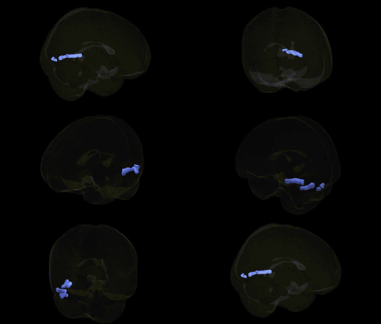
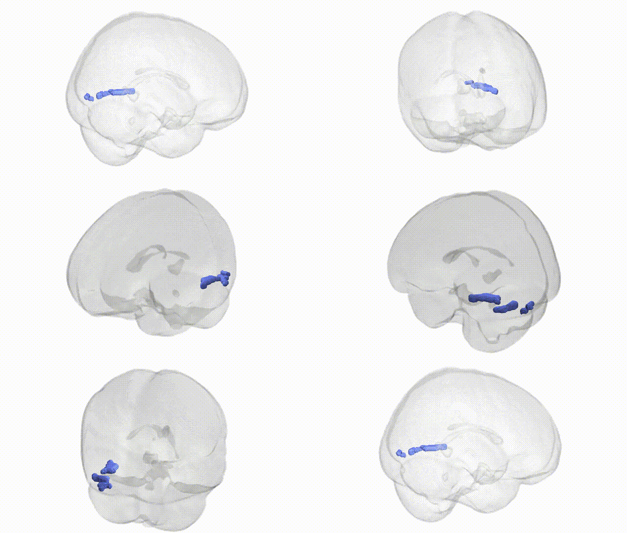
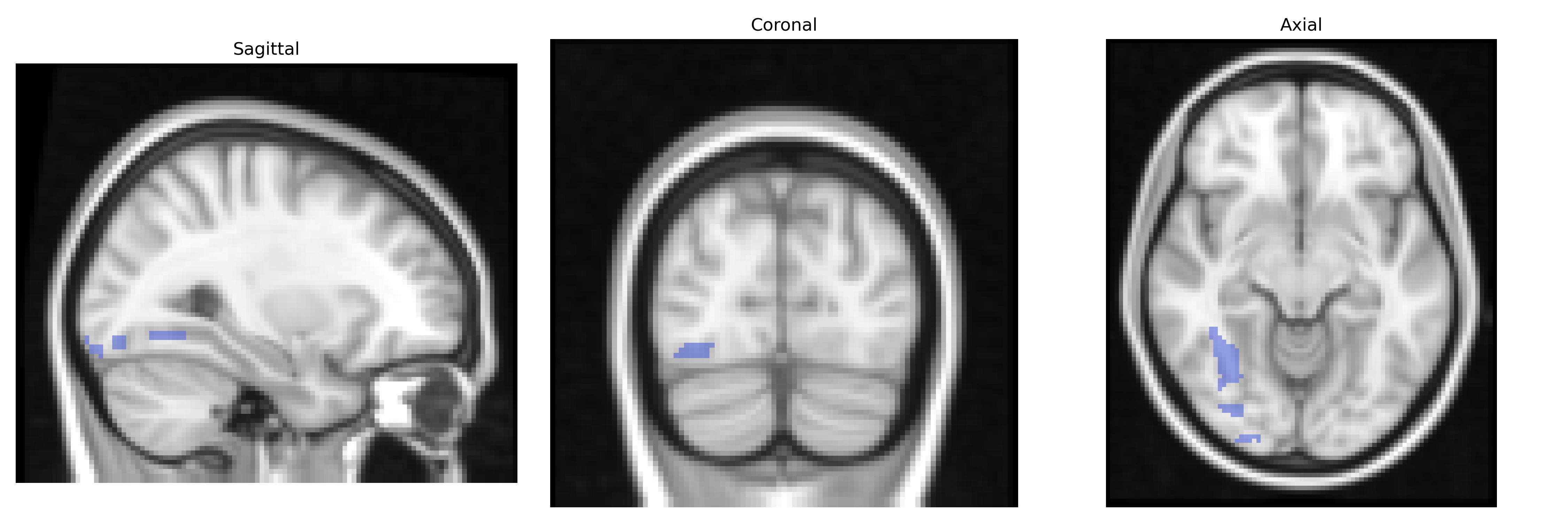
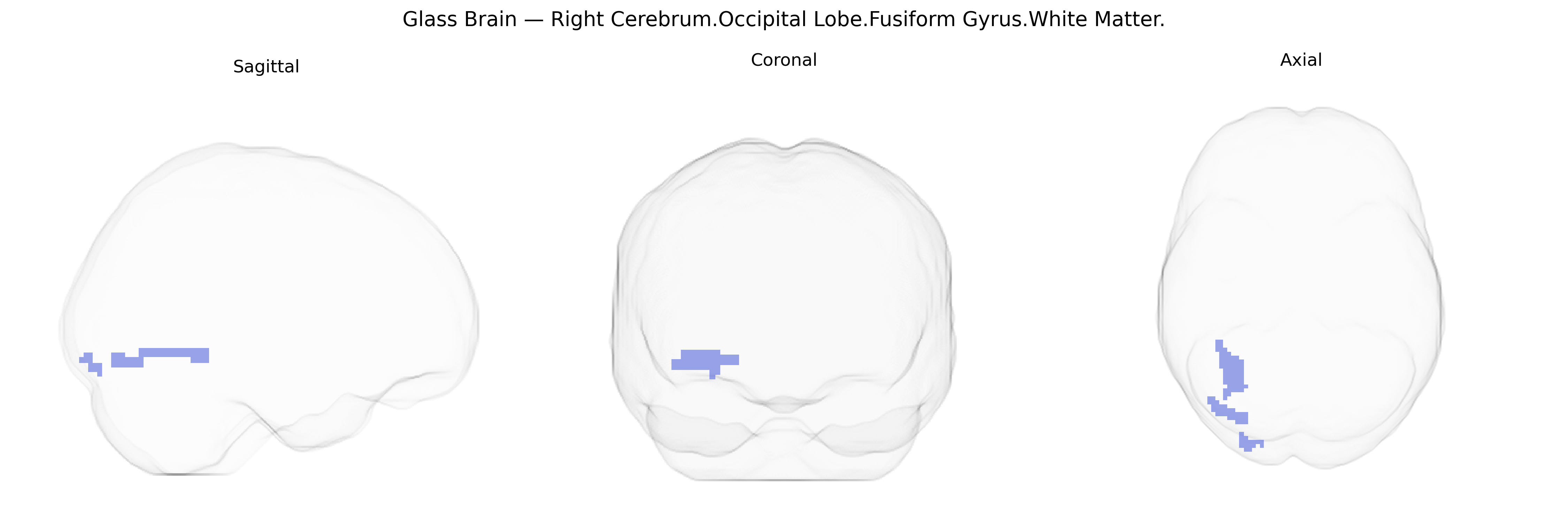
The bilateral Right Cerebrum.Occipital Lobe.Fusiform Gyrus.White Matter corresponds to the intra-gyral white matter tracts coursing within the fusiform gyrus of the occipital lobe, forming part of the ventral visual processing stream. This region contains association fibers linking occipital visual areas with more anterior temporal and parietal cortices and contributes to the integration and transmission of complex visual information, including object and face-related processing, toward higher-order recognition systems. Although the Talairach label specifies right occipital fusiform white matter, homologous pathways exist bilaterally and interconnect with long-range fasciculi such as the inferior longitudinal fasciculus and portions of the inferior fronto-occipital fasciculus, supporting perceptual integration, visual memory, and category-specific recognition in conjunction with adjacent gray matter structures. There is no direct Wikipedia article for the white matter of the occipital fusiform gyrus; a closely related structure is the fusiform gyrus: Fusiform gyrus.
The bilateral Right Cerebrum Occipital Lobe Fusiform Gyrus white matter, as defined in Talairach 2 mm space, corresponds to fiber pathways supporting the fusiform gyrus, a region central to high-level visual processing (including face, word, and object recognition), and has been indirectly implicated in genetic studies via imaging genetics and GWAS of brain structure and neuropsychiatric traits rather than through locus-specific analyses of this exact Talairach label. Large-scale GWAS of cortical surface area, thickness, and regional volumes (e.g., ENIGMA, UK Biobank) have identified common variants near genes involved in neurodevelopment (such as microtubule, synaptic, and axon guidance genes) that influence occipitotemporal and fusiform morphology and associated white matter microstructure, including loci near genes like DLG2, CNTNAP2, and others involved in synaptic organization and neuronal connectivity. Diffusion MRI GWAS have linked genetic variation affecting global and tract-specific white matter integrity (e.g., in genes related to myelination and oligodendrocyte function such as MAG, ERBB4, and others) to measures that partially overlap this occipitotemporal fusiform white matter zone. Functionally, genetic associations with disorders characterized by atypical fusiform structure or connectivity—such as autism spectrum disorder (ASD), developmental dyslexia, prosopagnosia, schizophrenia, and major depression—have implicated broad neurodevelopmental and synaptic gene sets through convergent evidence: ASD and dyslexia show altered fusiform activation and white matter organization, and GWAS/meta-analytic studies highlight genes involved in cortical patterning and connectivity that likely modulate these phenotypes. Additionally, polygenic risk for ASD, schizophrenia, and cognitive traits (e.g., educational attainment, general intelligence) has been associated with variation in occipitotemporal and fusiform cortical metrics, suggesting that the genetic architecture of these traits partly influences the development and integrity of the fusiform gyrus and its underlying white matter, though no single gene or variant can currently be said to specifically and uniquely target the bilateral Right Cerebrum Occipital Lobe Fusiform Gyrus white matter label in the Talairach 2 mm atlas.
Overview generated by GPT-4o (2026).
Region ID: 207
Hemisphere: bilateral
Atlas: Talairach labels 2mm
Full Quality Version: Download MP4
Full Quality Version: Download MP4
Wali Sidiqyar*, Gaurav Rudravaram*, Elyssa M. McMaster, Trent M. Schwartz, Adam M. Saunders, Kurt G. Schilling, Bennett A. Landman "Introducing SPINS: A Shared Public Visualization Library of Neuroanatomical Structures." Medical Imaging with Deep Learning- short paper
This resource is licensed under CC0 1.0 Universal (Public Domain).