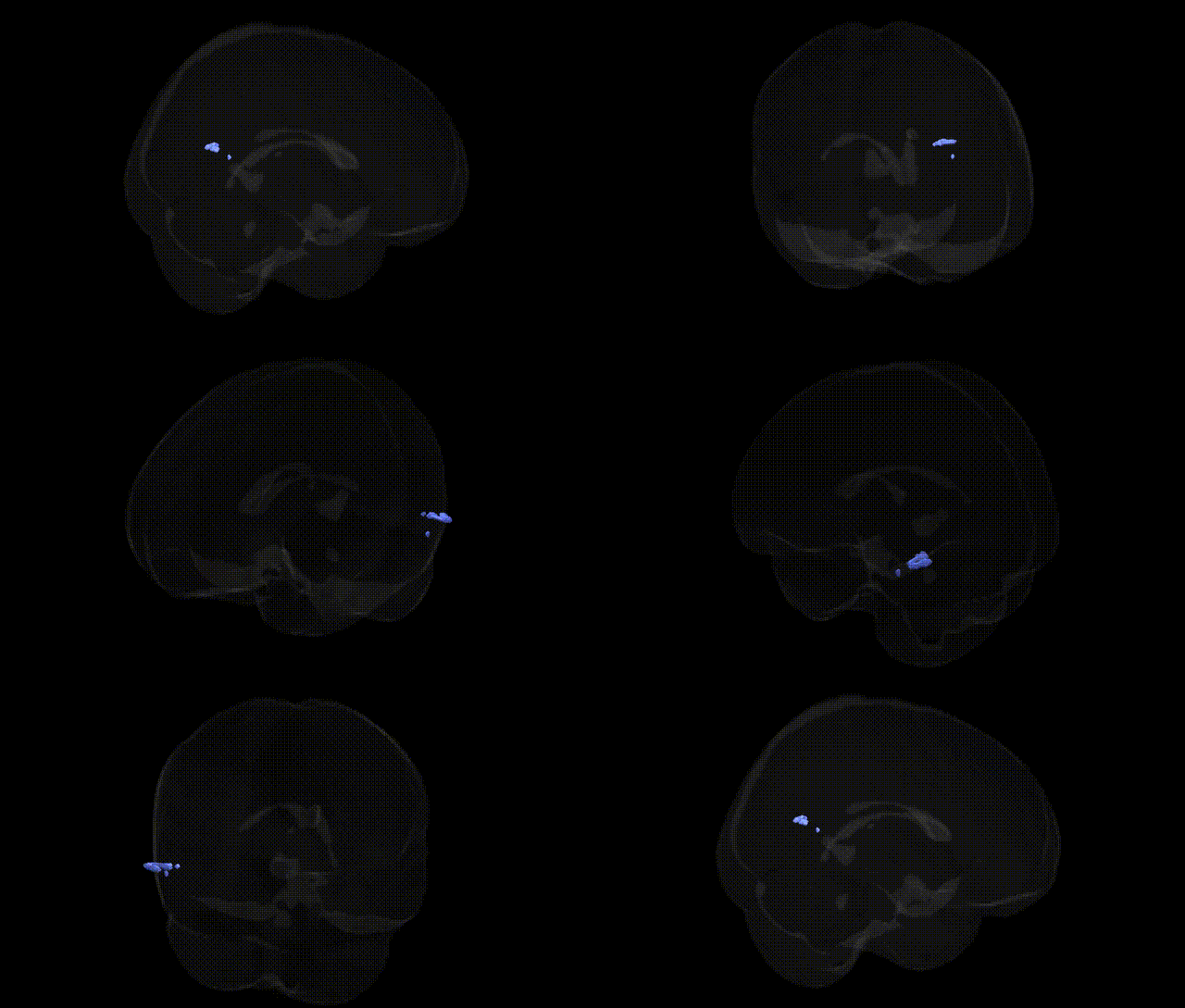
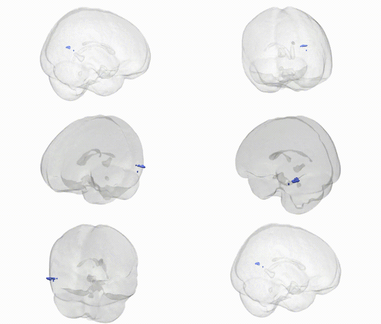
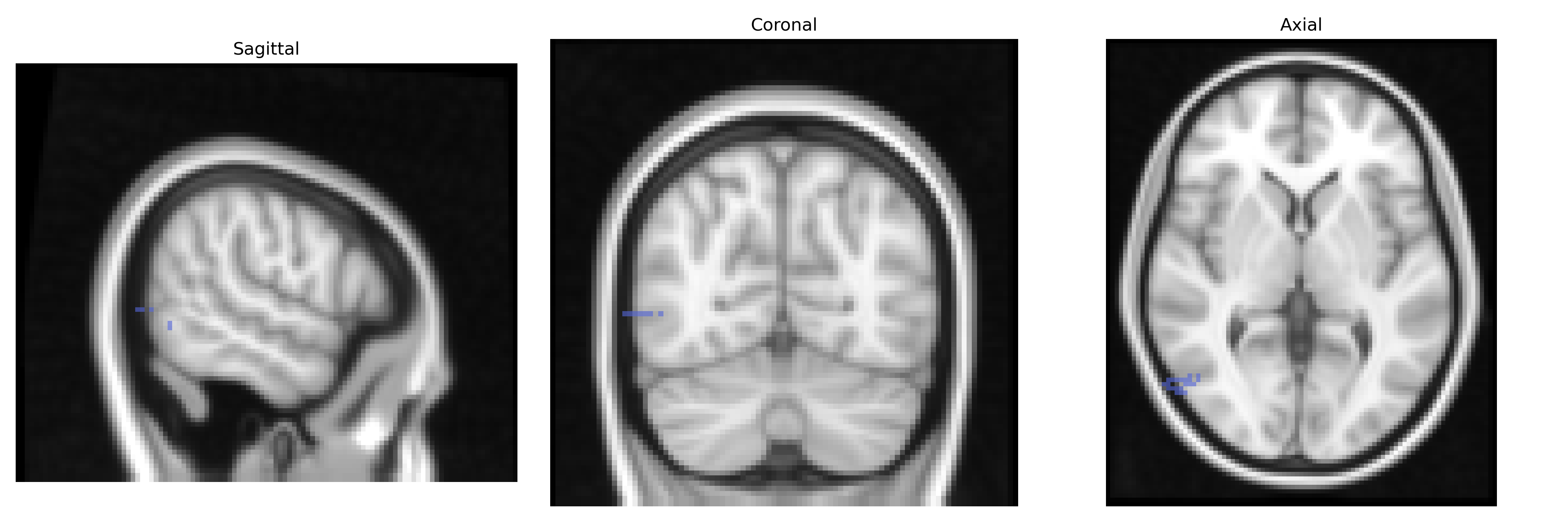
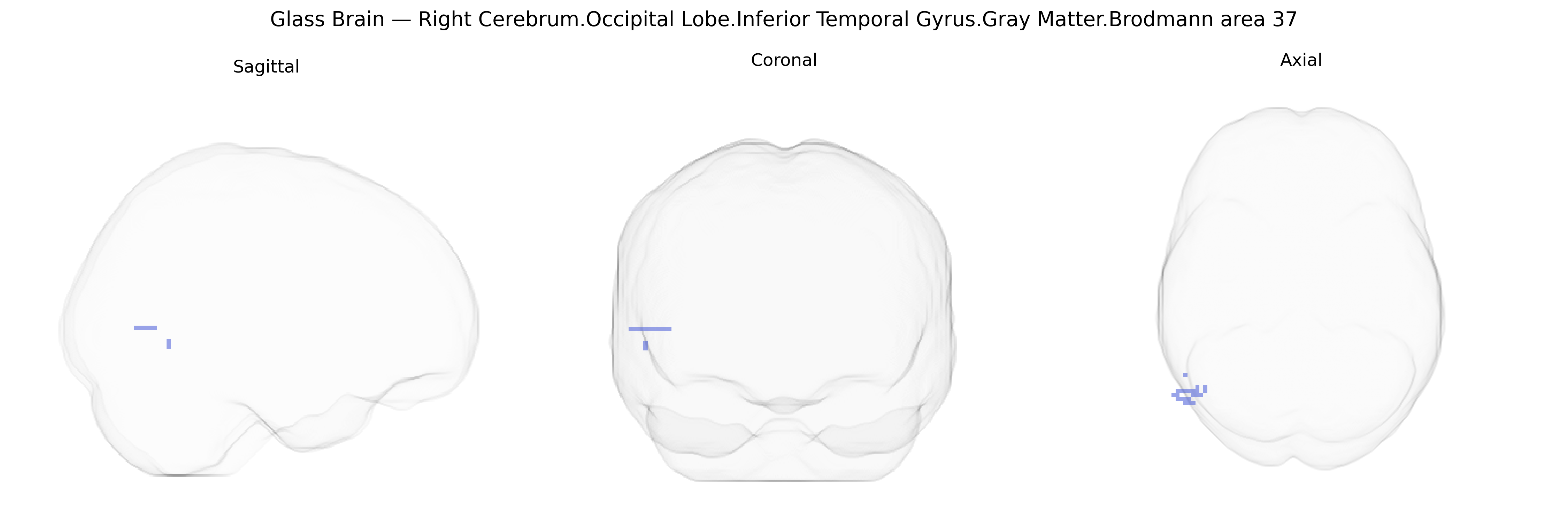
The bilateral Right Cerebrum.Occipital Lobe.Inferior Temporal Gyrus.Gray Matter.Brodmann area 37 corresponds to a portion of the ventral occipitotemporal cortex implicated in high-level visual processing, particularly in the recognition and categorization of complex visual stimuli such as faces, objects, and written words. Brodmann area 37 lies at the junction of the occipital and temporal lobes, encompassing parts of the fusiform and inferior temporal gyri, and forms a critical node within the ventral visual stream that transforms intermediate visual representations into semantic, language-related, and memory-linked information. This region exhibits strong connectivity with temporal, parietal, and frontal association cortices, supporting functions in multimodal integration, visual memory, and aspects of language comprehension, especially in relation to visually presented information. There is no direct link for “Brodmann area 37” as a standalone article; a closely related and overlapping structure is the Fusiform gyrus.
The bilateral right inferior temporal gyrus (ITG) gray matter in Brodmann area 37, a core component of the occipitotemporal cortex implicated in high-level visual processing and word/face recognition, is indirectly linked to several genetic associations through imaging genetics and GWAS of brain structure and function, though few studies target this precise Talairach-defined parcel. Large-scale GWAS on cortical thickness and surface area have implicated variants near genes such as KIAA0586, CENPW, and MIR9-2 in occipitotemporal and temporal cortical morphology, and polygenic influences on ITG/BA37 structure are evident in studies mapping distributed genetic effects on temporal and ventral visual stream regions. BA37 is consistently involved in reading and visual word-form processing, and genetic risk for dyslexia and language-related traits (e.g., variants in DCDC2, KIAA0319, ROBO1) has been associated with altered activation or structure in left occipitotemporal cortex, with some evidence for bilateral effects extending into inferior temporal regions. Face-processing networks including fusiform and adjacent BA37 territory show altered structure and activation in conditions with genetic underpinnings such as autism spectrum disorder and prosopagnosia; rare mutations (e.g., in MCTP2 in familial prosopagnosia) and common ASD risk variants have been linked to atypical ventral temporal circuitry encompassing BA37. GWAS of schizophrenia, bipolar disorder, and major depression have not identified BA37-specific loci, but imaging-genetic work indicates that risk variants (e.g., in CACNA1C, ZNF804A, and complement-related genes such as C4A) are associated with structural and functional alterations in temporal and occipitotemporal association cortices, including inferior temporal regions. Overall, genetic findings point to polygenic influences of neurodevelopmental, synaptic, and cell-adhesion genes on the structure and function of BA37 within broader temporal–occipitotemporal networks related to language, reading, face recognition, and social cognition, rather than discrete, region-exclusive GWAS signals for the specific Talairach 2 mm bilateral right BA37 label.
Overview generated by GPT-4o (2026).
Region ID: 422
Hemisphere: bilateral
Atlas: Talairach labels 2mm
Full Quality Version: Download MP4
Full Quality Version: Download MP4
Wali Sidiqyar*, Gaurav Rudravaram*, Elyssa M. McMaster, Trent M. Schwartz, Adam M. Saunders, Kurt G. Schilling, Bennett A. Landman "Introducing SPINS: A Shared Public Visualization Library of Neuroanatomical Structures." Medical Imaging with Deep Learning- short paper
This resource is licensed under CC0 1.0 Universal (Public Domain).