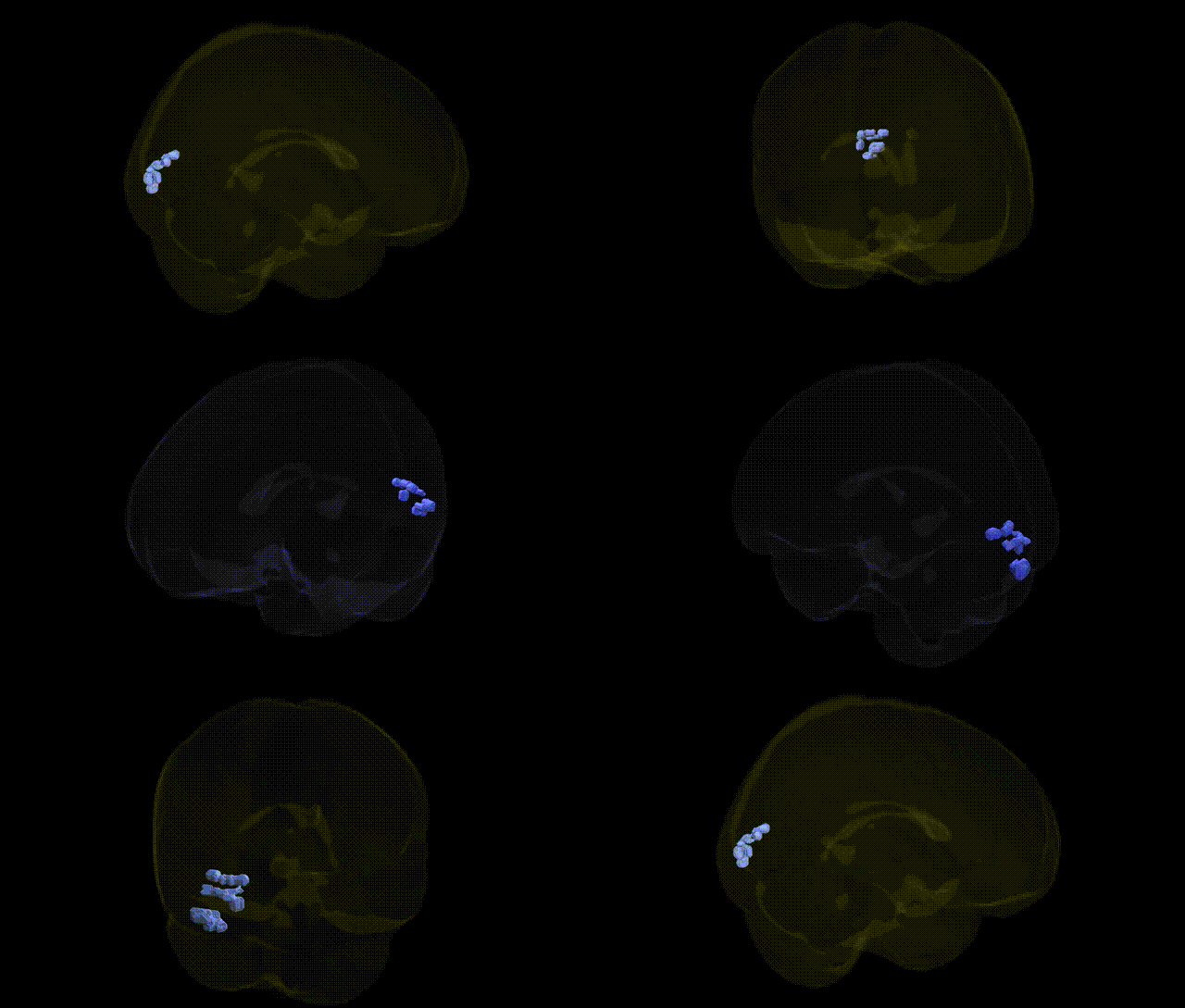
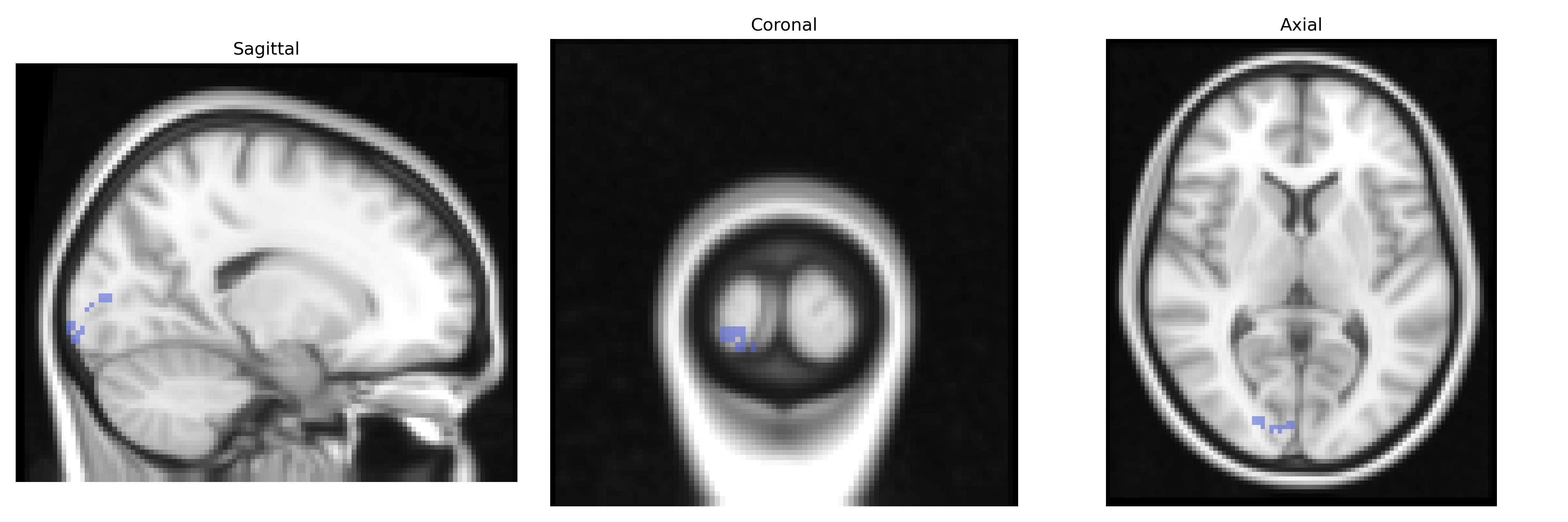
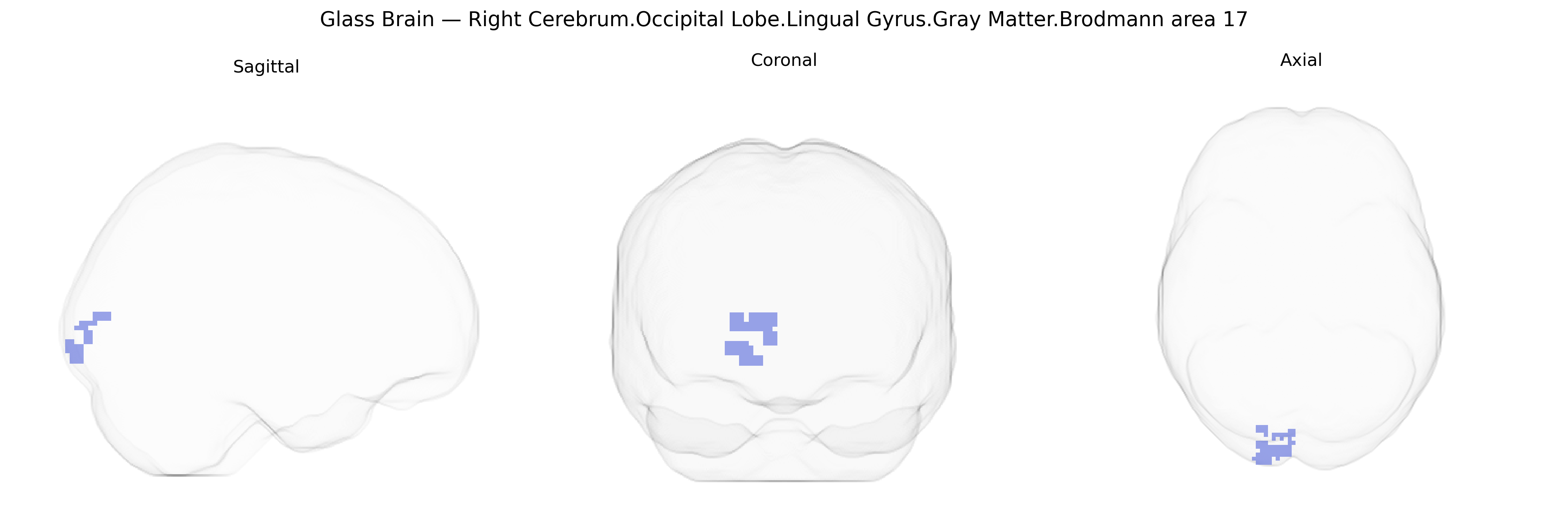
The bilateral Right Cerebrum.Occipital Lobe.Lingual Gyrus.Gray Matter.Brodmann area 17 corresponds to the primary visual cortex (V1) located along the calcarine fissure within the lingual gyrus of the occipital lobe, specialized for the initial cortical processing of visual information from the retina. This cytoarchitectonically defined region (BA17) receives highly organized input from the lateral geniculate nucleus of the thalamus and is characterized by a distinct laminar structure, including a prominent granular layer IV. Functionally, it encodes basic features of the visual scene such as orientation, spatial frequency, contrast, and ocular dominance, forming retinotopic maps that represent the visual field. The bilateral designation indicates homologous gray matter regions in both right and left hemispheres that together support conscious visual perception and serve as the gateway for subsequent visual processing in extrastriate areas.
The bilateral lingual gyrus gray matter in Brodmann area 17 (primary visual cortex) has been implicated in multiple genetic and GWAS-based associations, largely through imaging genetics and brain-structure GWAS rather than region-specific candidate gene studies. Large-scale neuroimaging GWAS (e.g., ENIGMA, UK Biobank) have identified common variants affecting occipital and visual cortical thickness, surface area, and volume, with signals enriched near genes involved in neurodevelopment, synaptic function, and axon guidance (such as variants near PAX6, FGFR genes, and other regulators of cortical patterning), though individual loci specific to the lingual gyrus per se are rarely isolated. Functional imaging genetics studies link polymorphisms in genes such as BDNF (e.g., Val66Met) and dopaminergic or serotonergic genes to altered activation or connectivity in visual cortex during tasks involving visual memory, emotional face processing, or visual attention. In psychiatric and neurodevelopmental disorders, structural and functional alterations of the lingual gyrus and adjacent BA17 regions have been reported in schizophrenia, major depressive disorder, bipolar disorder, autism spectrum disorder, and dyslexia, where GWAS-implicated risk genes (including those influencing synaptic plasticity, glutamatergic signaling, and cortical development) are thought to contribute to abnormal occipital/visual network organization rather than exclusively targeting this region. GWAS of migraine, particularly visual aura, and of photic epilepsy/visual sensitivity indicate that visual cortex excitability is partly genetically driven, with implicated loci in ion-channel and neurotransmission genes plausibly affecting BA17 circuitry. Finally, polygenic scores for cognitive traits such as intelligence, educational attainment, and reading ability show associations with structural metrics in occipital cortex, including lingual gyrus gray matter, reinforcing that genetic influences on neurocognitive function extend into primary visual regions and their integration with higher-order networks.
Overview generated by GPT-4o (2026).
Region ID: 245
Hemisphere: bilateral
Atlas: Talairach labels 2mm
Full Quality Version: Download MP4
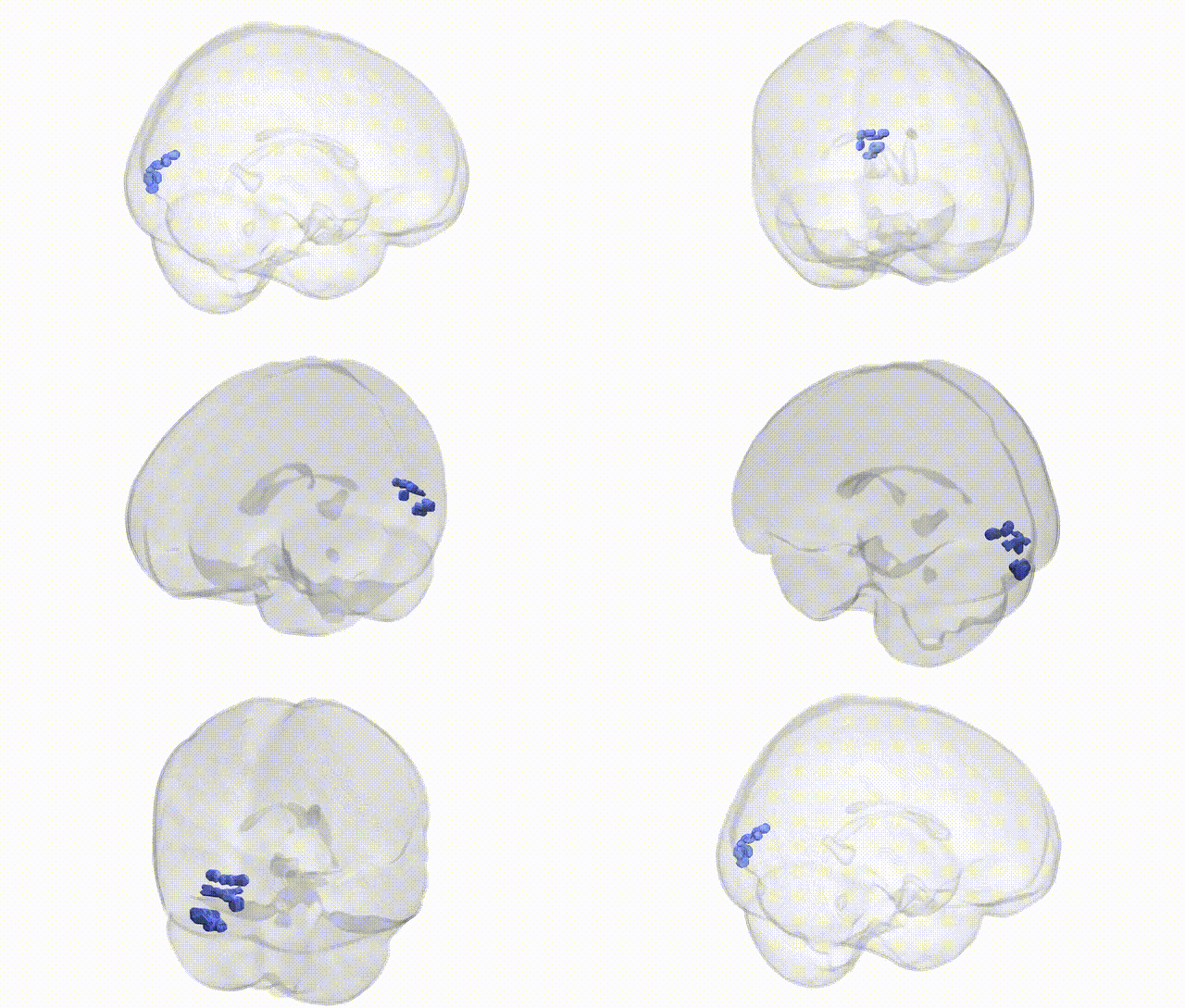
Full Quality Version: Download MP4
Wali Sidiqyar*, Gaurav Rudravaram*, Elyssa M. McMaster, Trent M. Schwartz, Adam M. Saunders, Kurt G. Schilling, Bennett A. Landman "Introducing SPINS: A Shared Public Visualization Library of Neuroanatomical Structures." Medical Imaging with Deep Learning- short paper
This resource is licensed under CC0 1.0 Universal (Public Domain).