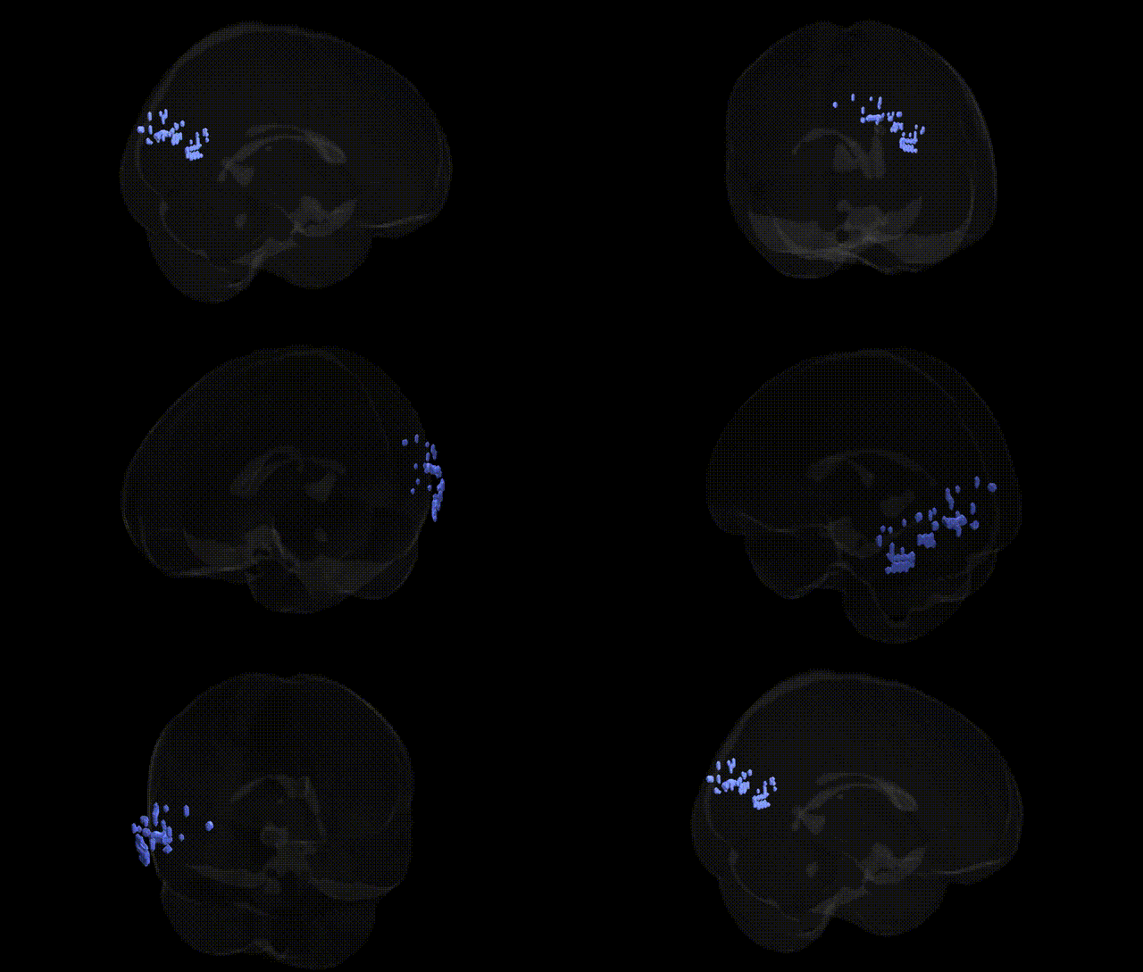
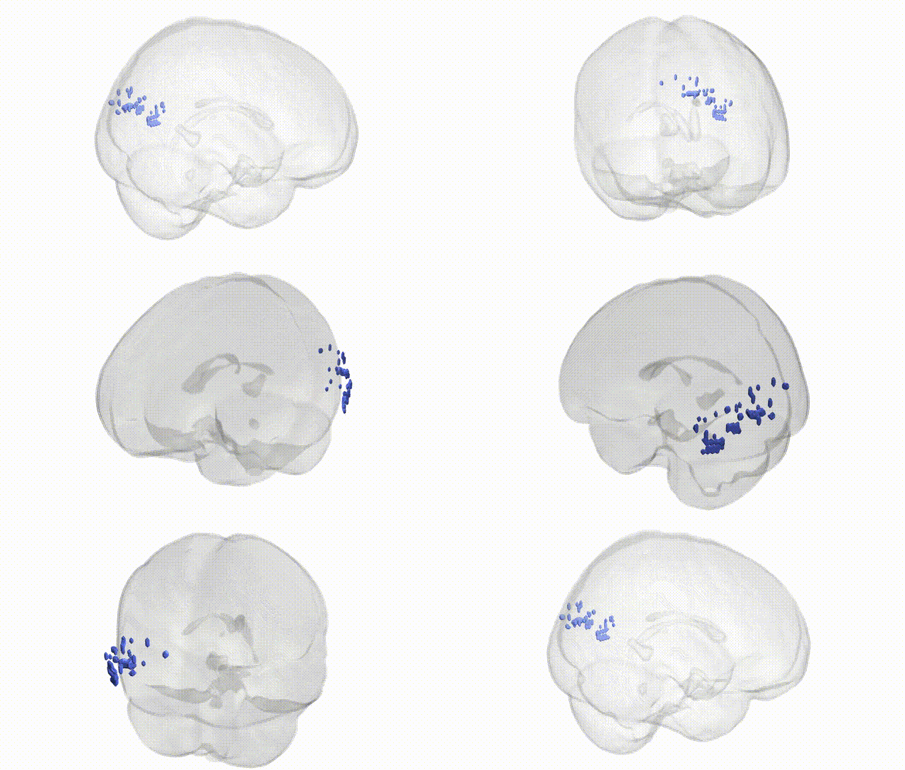
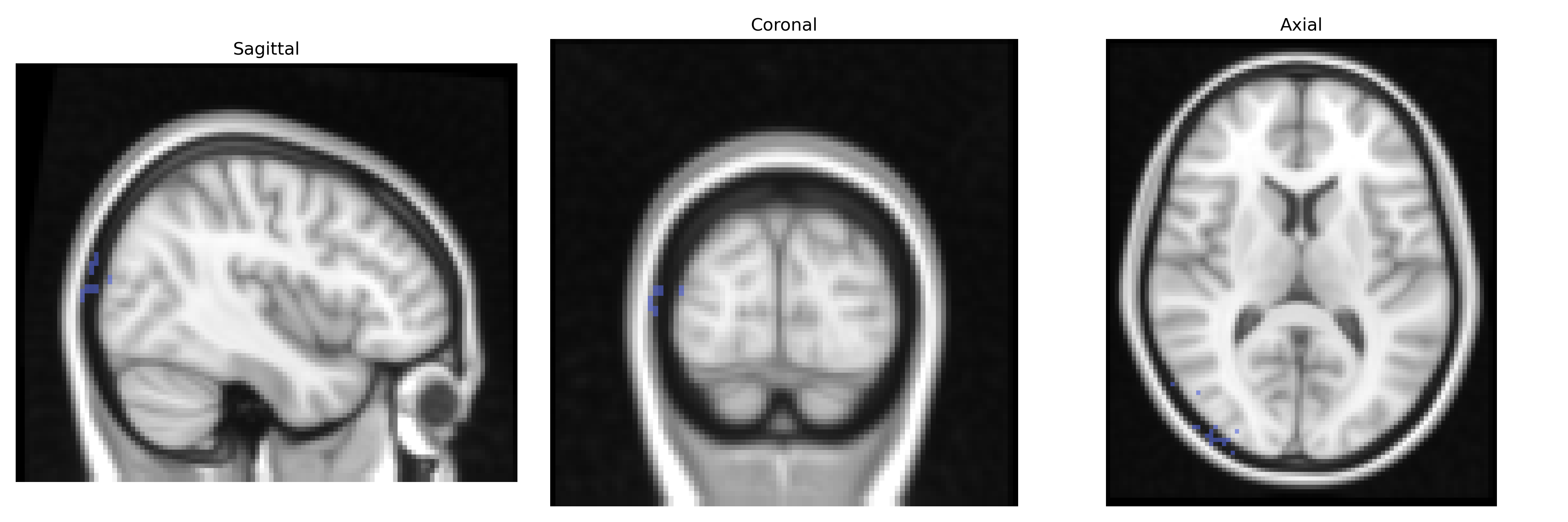
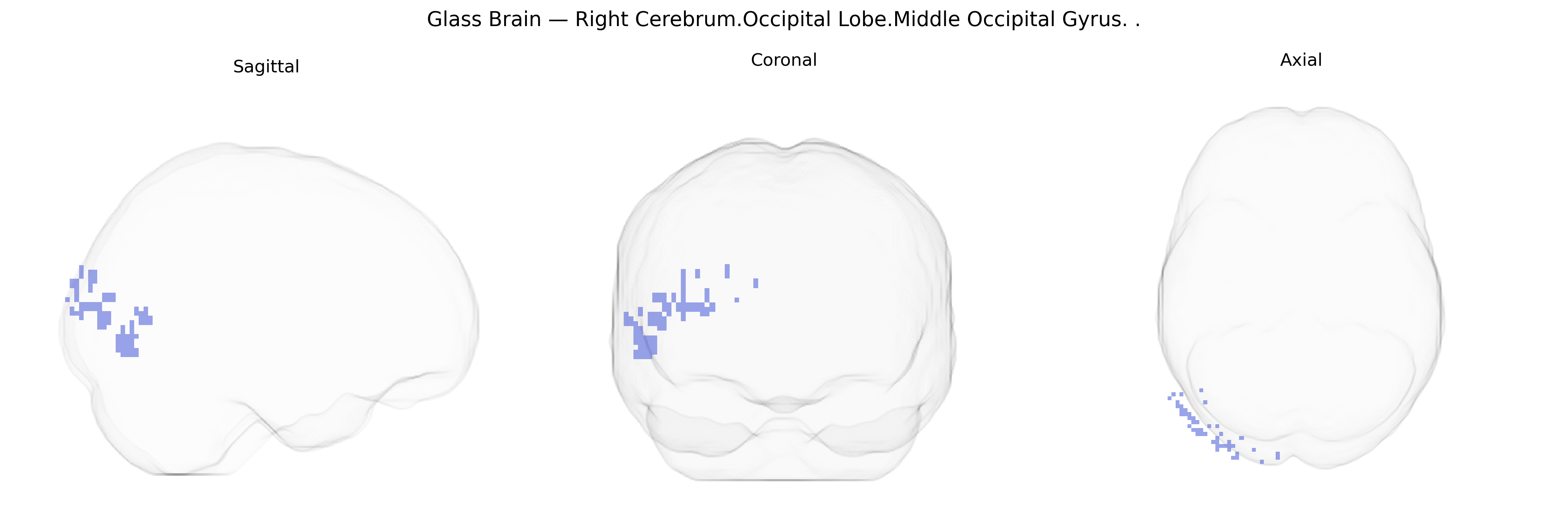
The bilateral Right Cerebrum.Occipital Lobe.Middle Occipital Gyrus corresponds to cortex in the middle portion of the occipital lobe, primarily encompassing parts of Brodmann areas 18 and 19, which form secondary and associative visual regions. This gyrus participates in the processing of complex visual information, including object form, motion, and spatial relationships, and serves as an important relay between primary visual cortex (V1) and higher-order visual areas in the temporal and parietal lobes. It contributes to visual perception, recognition, and integration of visual stimuli into broader cognitive representations, with functional asymmetries modulated by hemispheric specialization and task demands. There is no direct link for the middle occipital gyrus; a closely related structure is the Occipital lobe.
The bilateral Right Cerebrum Occipital Lobe Middle Occipital Gyrus, a core visual association area, has been implicated in genetic studies primarily through imaging genetics and GWAS of brain structure and function rather than region-specific locus mapping in the Talairach 2 mm atlas. Variants in genes involved in synaptic plasticity, neurodevelopment, and visual pathways—such as BDNF, COMT, GRIN2B, and CACNA1C—have been associated with occipital cortical thickness, surface area, and functional activation during visual processing, with some studies showing that risk alleles for schizophrenia, bipolar disorder, and major depression modulate activation or connectivity in middle occipital regions during visual or emotional tasks. Large GWAS of brain MRI measures (e.g., ENIGMA and UK Biobank) have identified multiple loci influencing occipital cortical morphology and visual network connectivity, including variants near genes such as HMGA2, CENPW, IGF1, and others that broadly affect cortical development, though these are typically reported at the lobe or visual-network level rather than specifically the middle occipital gyrus. Occipital regions including the middle occipital gyrus show altered structure and function in genetically influenced disorders such as schizophrenia, autism spectrum disorder, ADHD, and migraine with aura, and polygenic risk scores for these conditions have been linked to variance in occipital cortical metrics. Overall, genetic associations with this Talairach-defined region are indirect, arising from broader visual cortex, occipital lobe, and brain-wide GWAS findings that implicate neurodevelopmental, synaptic, and vascular genes in shaping its anatomy and activity.
Overview generated by GPT-4o (2026).
Region ID: 311
Hemisphere: bilateral
Atlas: Talairach labels 2mm
Full Quality Version: Download MP4
Full Quality Version: Download MP4
Wali Sidiqyar*, Gaurav Rudravaram*, Elyssa M. McMaster, Trent M. Schwartz, Adam M. Saunders, Kurt G. Schilling, Bennett A. Landman "Introducing SPINS: A Shared Public Visualization Library of Neuroanatomical Structures." Medical Imaging with Deep Learning- short paper
This resource is licensed under CC0 1.0 Universal (Public Domain).