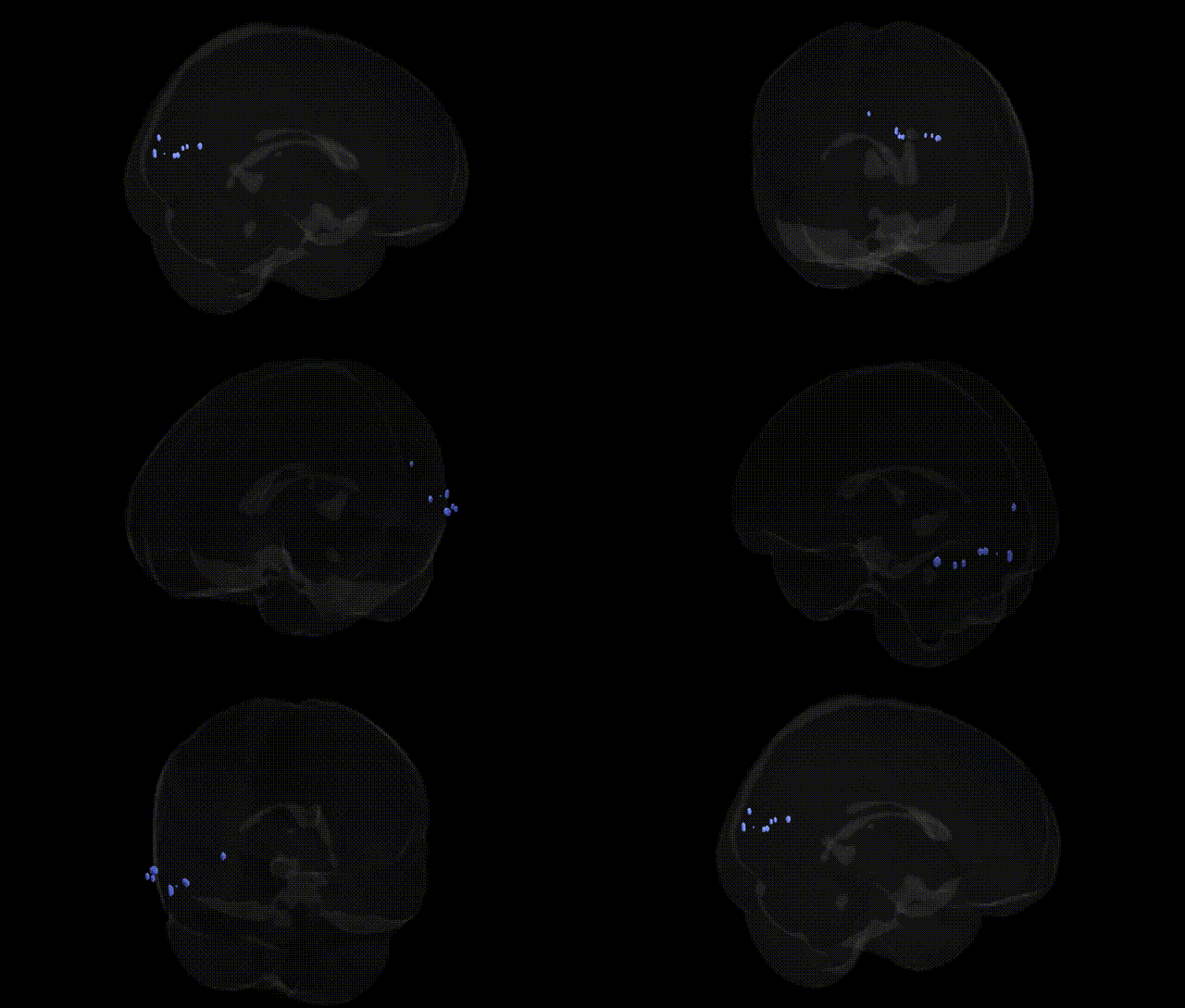
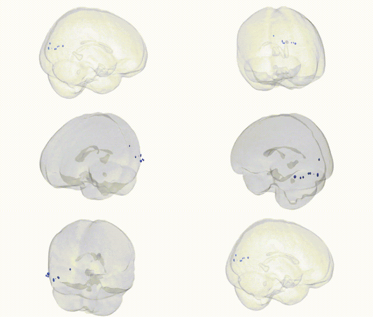
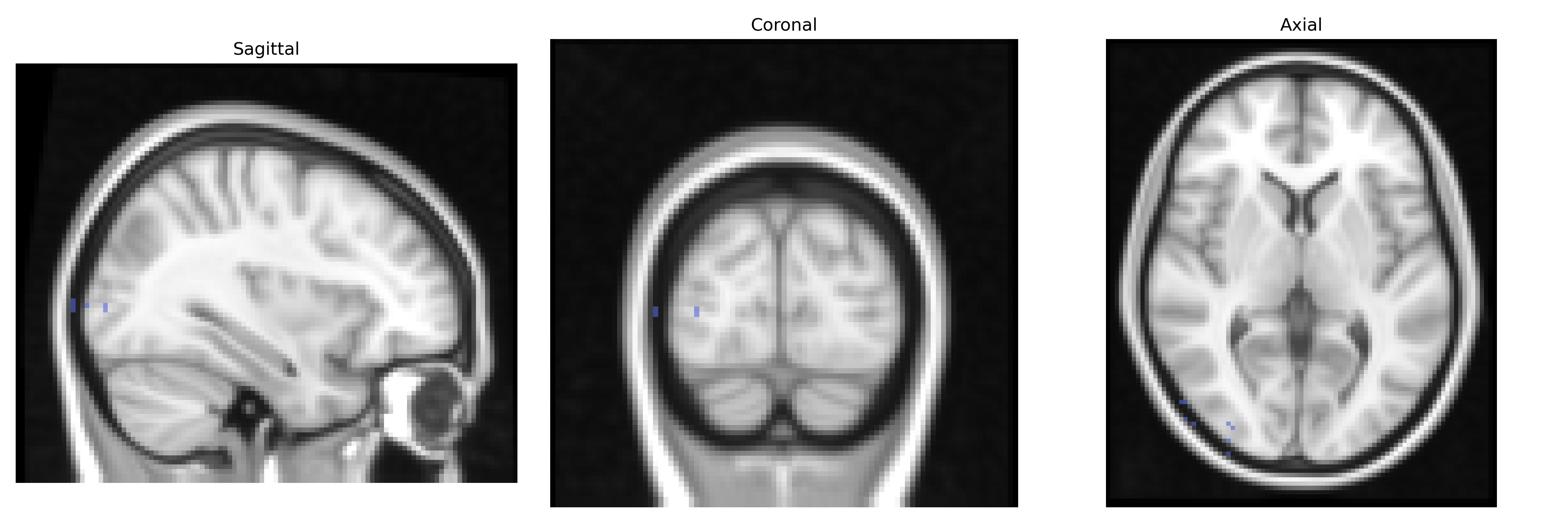
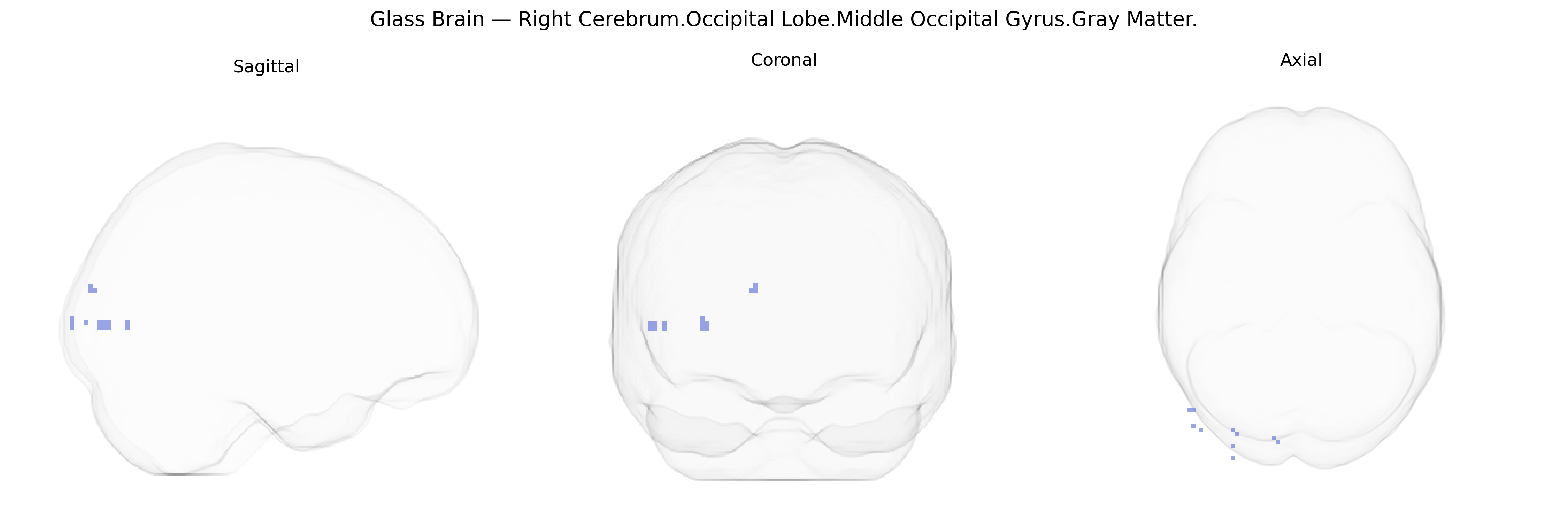
The bilateral Right Cerebrum.Occipital Lobe.Middle Occipital Gyrus.Gray Matter corresponds to cortical tissue in the middle segment of the occipital lobe, primarily involved in intermediate to high-level visual processing. This region contributes to the analysis of object form, motion, and spatial relationships, integrating visual inputs from primary visual cortex and relaying processed information to dorsal and ventral visual pathways. Neuronal populations in the middle occipital gyrus participate in visuospatial attention, perception of complex scenes, and aspects of visually guided behavior, and they interact with parietal and temporal cortices for multimodal integration. There is no direct link, but it is part of the occipital lobe: Occipital lobe.
The bilateral Right Cerebrum.Occipital Lobe.Middle Occipital Gyrus.Gray Matter, as defined in the Talairach 2 mm atlas, falls within the lateral occipital cortex implicated in visual processing, object recognition, and higher-order perceptual integration, and has been repeatedly highlighted in imaging genetics and GWAS of brain structure. Large-scale neuroimaging GWAS (e.g., ENIGMA and UK Biobank cohorts) have identified common variants in genes involved in neurodevelopment and synaptic function—such as HMGA2, IGF1, and multiple loci near MAPT and BDNF—that associate with occipital and lateral occipital cortical thickness, surface area, and gray matter volume, although most signals are not specific to the middle occipital gyrus alone. Polygenic influences from general cognitive ability, educational attainment, and schizophrenia risk scores have been linked to variation in occipital gray matter, including middle/lateral occipital regions, consistent with their role in visual cognition and higher-order processing. Occipital gyrus morphology has also been associated with genetic risk for neurodevelopmental and psychiatric disorders, such as autism spectrum disorder, major depression, and bipolar disorder, in which GWAS-identified risk variants and polygenic scores correlate with altered occipital cortical structure or activation patterns. Additionally, variants implicated in migraine, visual aura, and occipital epilepsy have been indirectly connected to functional alterations in occipital cortex, including the middle occipital gyrus, although precise gyrus-level genetic localization remains limited. Overall, genetic studies support a highly polygenic architecture affecting gray matter in this region, with overlapping loci contributing to visual processing, cognitive traits, and vulnerability to neuropsychiatric and neurological conditions.
Overview generated by GPT-4o (2026).
Region ID: 521
Hemisphere: bilateral
Atlas: Talairach labels 2mm
Full Quality Version: Download MP4
Full Quality Version: Download MP4
Wali Sidiqyar*, Gaurav Rudravaram*, Elyssa M. McMaster, Trent M. Schwartz, Adam M. Saunders, Kurt G. Schilling, Bennett A. Landman "Introducing SPINS: A Shared Public Visualization Library of Neuroanatomical Structures." Medical Imaging with Deep Learning- short paper
This resource is licensed under CC0 1.0 Universal (Public Domain).