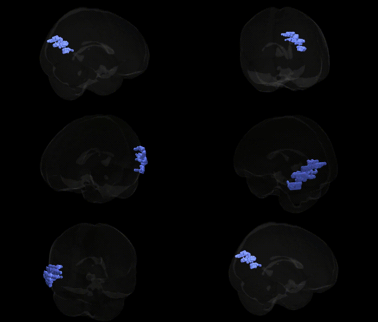
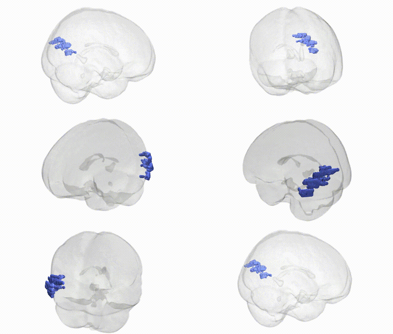
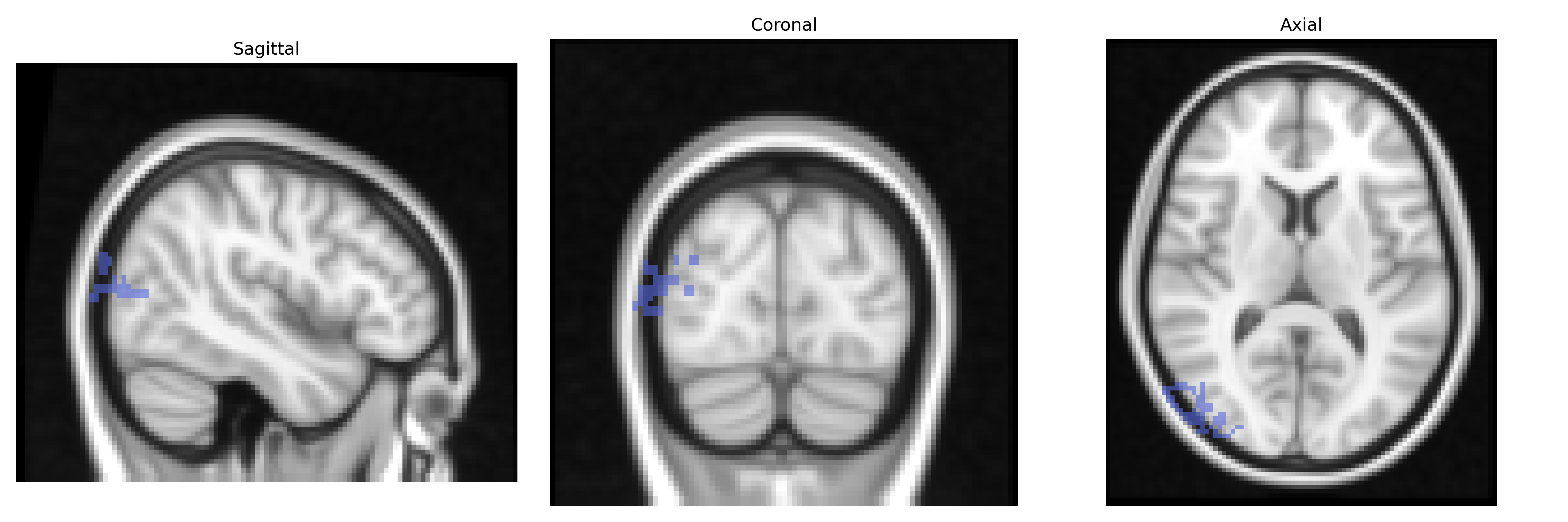
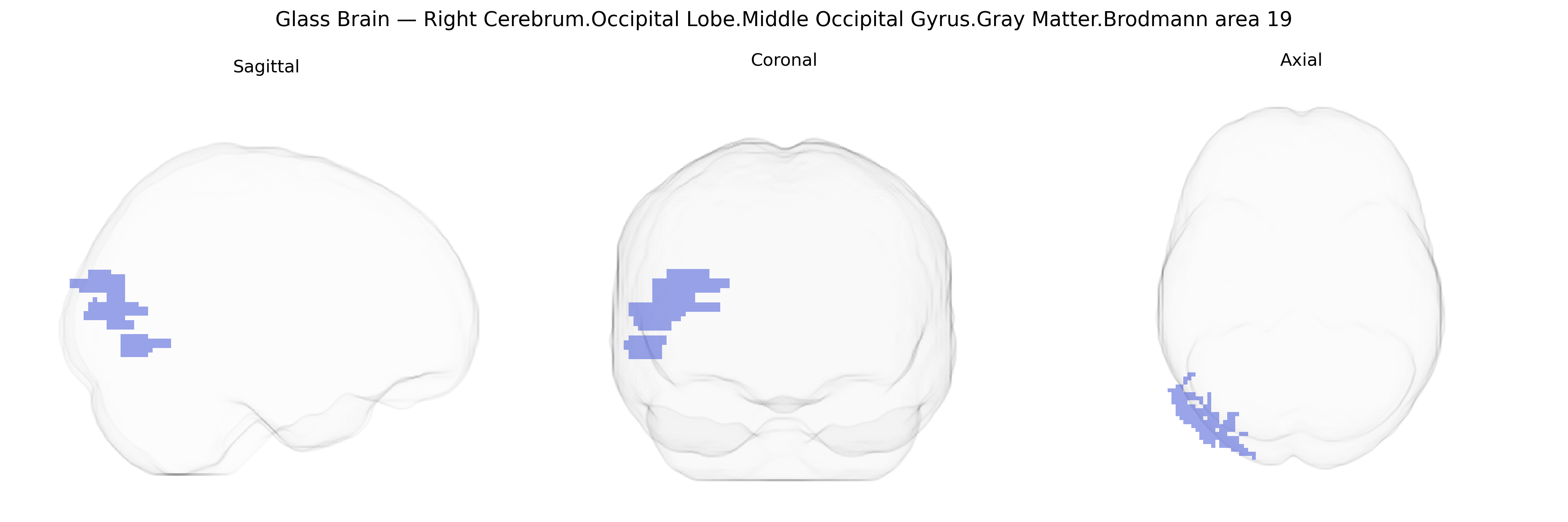
The bilateral Right Cerebrum.Occipital Lobe.Middle Occipital Gyrus.Gray Matter.Brodmann area 19 corresponds to associative visual cortex within the occipital lobe, situated lateral to primary and secondary visual areas (BA17 and BA18). This region participates in higher-order processing of visual information, including integration of complex shapes, motion, and spatial relationships, and contributes to visual recognition by relaying and transforming inputs from earlier visual areas to temporal and parietal cortices. Neuronal populations in BA19 are involved in constructing representations necessary for object perception, visuospatial analysis, and coordination of visual information with other sensory and cognitive systems. There is no direct link for this exact composite label; a closely related area is Brodmann area 19.
The bilateral right middle occipital gyrus in Brodmann area 19, a key part of the extrastriate visual cortex implicated in higher-order visual processing, has been indirectly linked to several genetic findings through imaging genetics and GWAS, although few studies target this specific Talairach-defined parcel. Variants in genes related to synaptic plasticity and neurodevelopment—such as BDNF (e.g., Val66Met), NRG1, and CACNA1C—have been associated with structural and functional differences in occipital and associative visual cortices, including BA19, in contexts of schizophrenia, bipolar disorder, and major depression. Visual cortical regions encompassing BA19 have shown volume or thickness alterations tied to common risk alleles from large psychiatric GWAS (e.g., genome-wide polygenic risk scores for schizophrenia and autism spectrum disorder), suggesting genetically influenced vulnerability of higher-order visual areas in these conditions. GWAS of brain imaging phenotypes (e.g., UK Biobank and ENIGMA consortia) have identified loci affecting occipital cortical surface area and thickness—often near genes involved in neuronal migration and axon guidance such as DCC and ROBO family members—though these findings typically reference broader occipital regions rather than BA19 specifically. In addition, genetic variants contributing to visual traits (myopia, macular degeneration, and processing speed) and reading-related disorders (such as dyslexia, with genes like KIAA0319 and DCDC2) have been associated with altered activation and connectivity within occipital and temporo-occipital cortices that include BA19. Overall, the genetic associations for this precise bilateral right BA19 region are inferred largely from broader occipital or visual-association cortex analyses, with converging evidence that common neurodevelopmental and synaptic genes modulate its structure and function in relation to psychiatric, cognitive, and visual phenotypes.
Overview generated by GPT-4o (2026).
Region ID: 316
Hemisphere: bilateral
Atlas: Talairach labels 2mm
Full Quality Version: Download MP4
Full Quality Version: Download MP4
Wali Sidiqyar*, Gaurav Rudravaram*, Elyssa M. McMaster, Trent M. Schwartz, Adam M. Saunders, Kurt G. Schilling, Bennett A. Landman "Introducing SPINS: A Shared Public Visualization Library of Neuroanatomical Structures." Medical Imaging with Deep Learning- short paper
This resource is licensed under CC0 1.0 Universal (Public Domain).