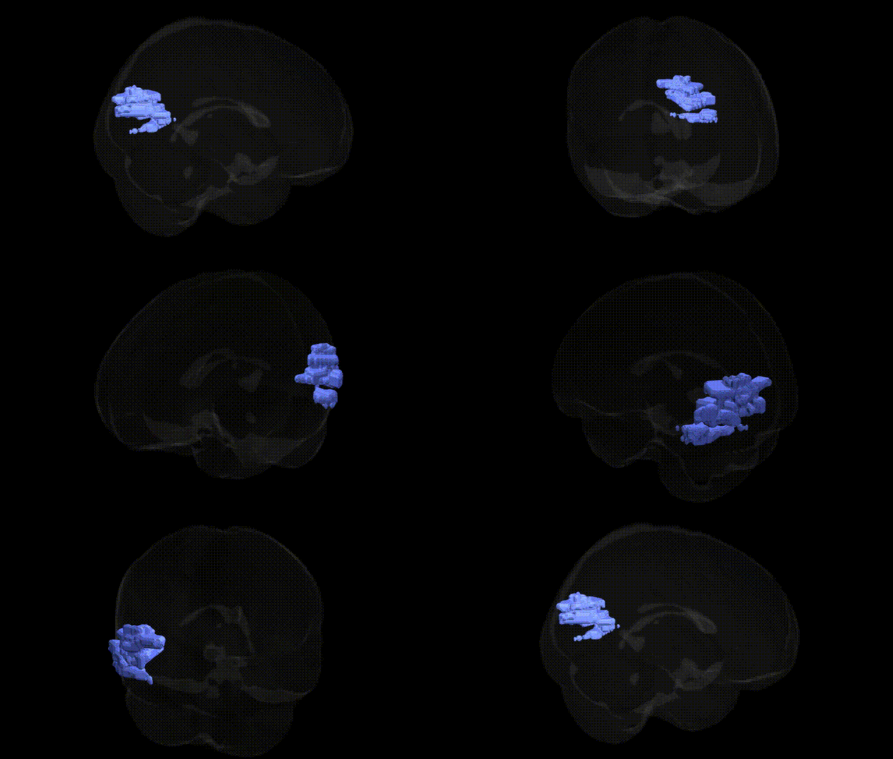
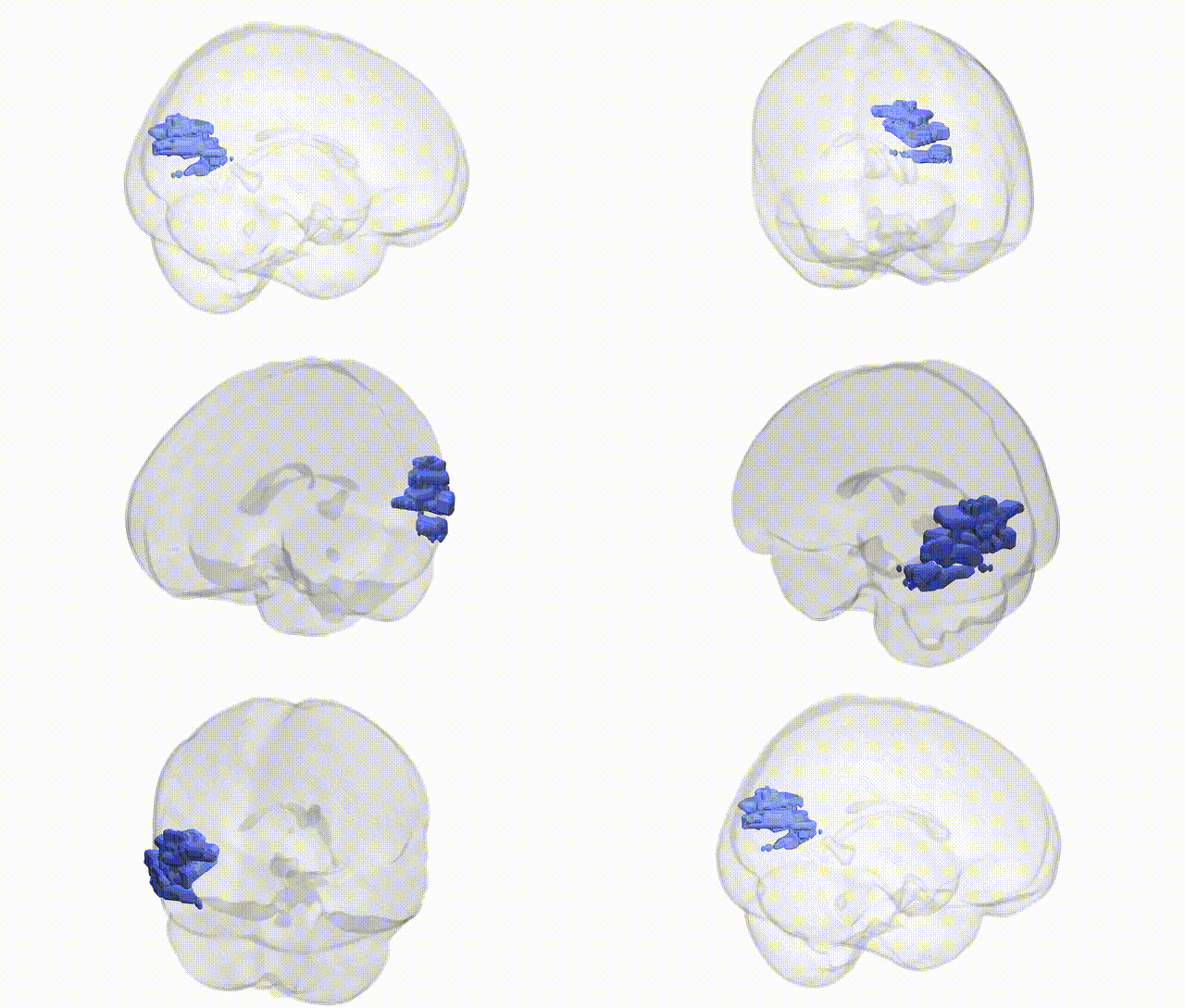
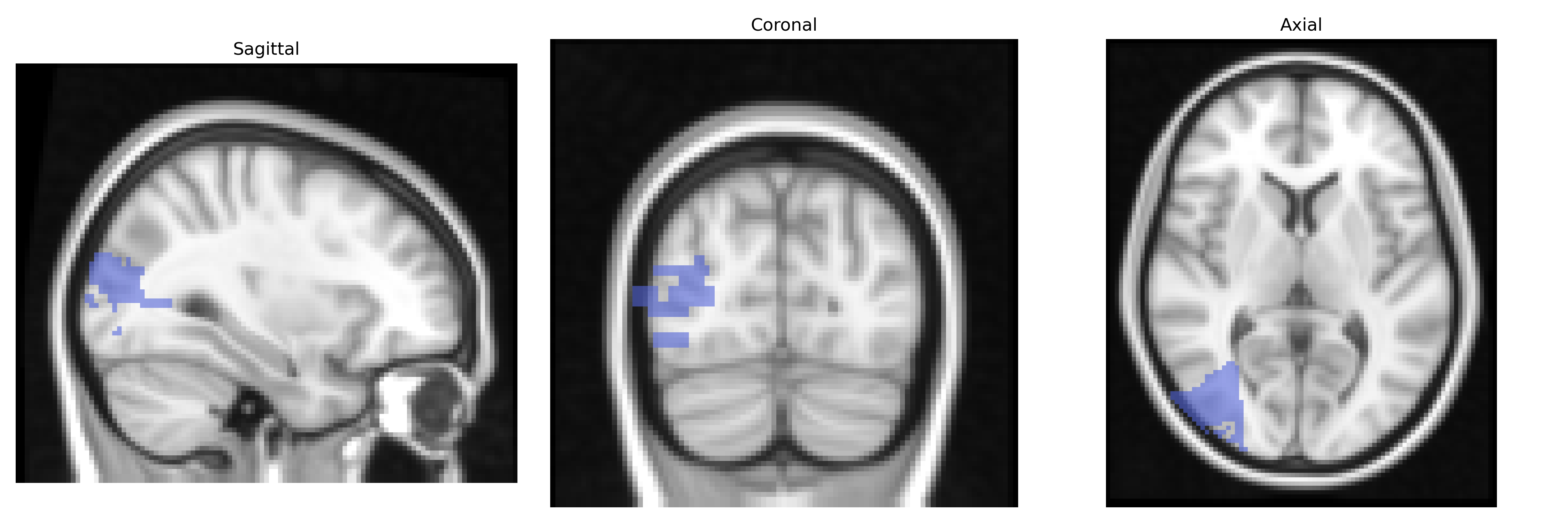
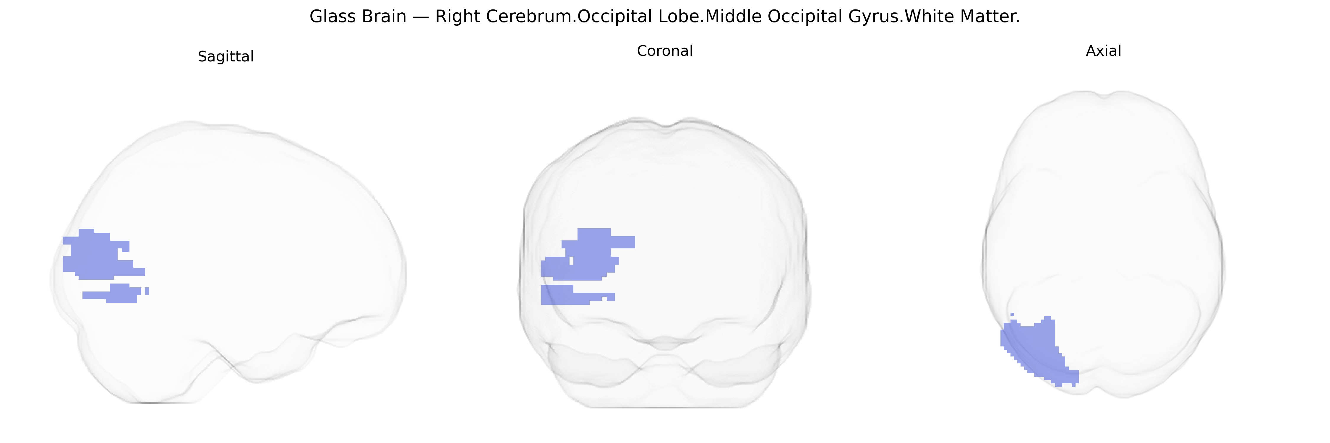
The bilateral Right Cerebrum.Occipital Lobe.Middle Occipital Gyrus.White Matter corresponds to the subcortical fiber systems underlying the middle occipital gyrus in the occipital lobe, encompassing association and projection pathways that support visual information processing. This white matter region carries axons interconnecting occipital visual areas with temporal and parietal cortices, contributing to the integration of visual features with spatial, motion, and higher-order perceptual functions. Although the Talairach label specifies the right cerebrum, the term “bilateral” indicates homologous tracts in both hemispheres, reflecting the generally symmetrical organization of occipital white matter. There is no direct Wikipedia article for this specific white matter subdivision; a related cortical structure is the Middle occipital gyrus.
The bilateral Right Cerebrum Occipital Lobe Middle Occipital Gyrus White Matter, as defined in the Talairach 2 mm atlas, lies within the broader occipital and extrastriate visual networks that have been repeatedly implicated in imaging genetics and GWAS of brain structure and function, although few studies isolate this tract-specific parcel. Large-scale neuroimaging GWAS (e.g., ENIGMA, UK Biobank) have identified multiple loci influencing global and regional occipital lobe volume, cortical thickness, and white matter microstructure (fractional anisotropy, mean diffusivity), with notable contributors including variants near genes involved in neurodevelopment and axonal guidance such as HMGA2, IGF1, and cell-adhesion and synaptic genes (e.g., DCC, NCAM1, CNTN4), which affect occipital white matter integrity. Visual cortex and occipital white matter measures overlapping Middle Occipital Gyrus territory have been associated with polygenic influences on intelligence, educational attainment, and processing speed, as well as with neurodevelopmental and psychiatric conditions including autism spectrum disorder, schizophrenia, and ADHD, where risk alleles in synaptic and neurodevelopmental pathways show downstream effects on occipital connectivity and structure. Occipital white matter tracts passing through or near this region (such as portions of the inferior longitudinal fasciculus and inferior fronto-occipital fasciculus) also show genetic correlations with reading ability and dyslexia, migraine and visual aura, and disorders of visual perception, reflecting shared polygenic architectures that modulate visual information transfer. Overall, genetic studies support a highly polygenic, pleiotropic architecture in which risk and trait-associated variants for cognition, neurodevelopmental disorders, and sensory phenotypes exert measurable effects on occipital white matter—including the Middle Occipital Gyrus segment—through pathways regulating cortical development, myelination, and long-range connectivity.
Overview generated by GPT-4o (2026).
Region ID: 309
Hemisphere: bilateral
Atlas: Talairach labels 2mm
Full Quality Version: Download MP4
Full Quality Version: Download MP4
Wali Sidiqyar*, Gaurav Rudravaram*, Elyssa M. McMaster, Trent M. Schwartz, Adam M. Saunders, Kurt G. Schilling, Bennett A. Landman "Introducing SPINS: A Shared Public Visualization Library of Neuroanatomical Structures." Medical Imaging with Deep Learning- short paper
This resource is licensed under CC0 1.0 Universal (Public Domain).