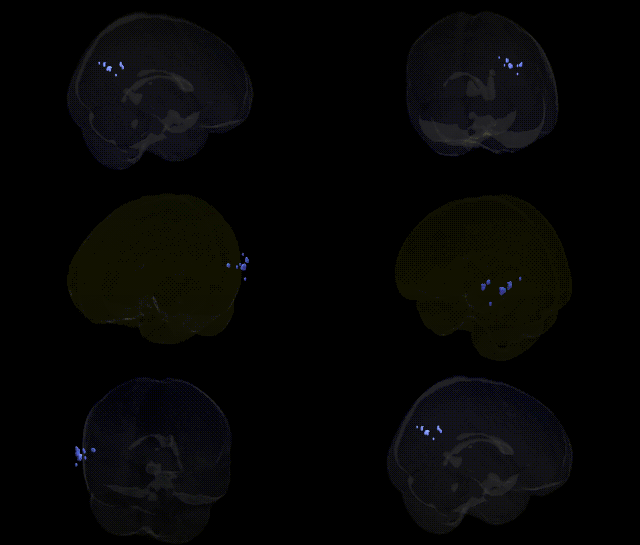
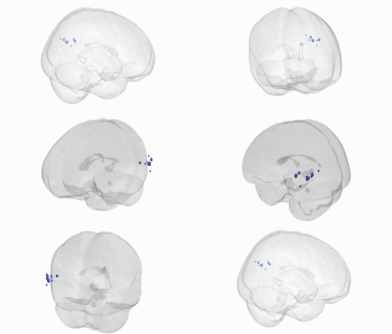
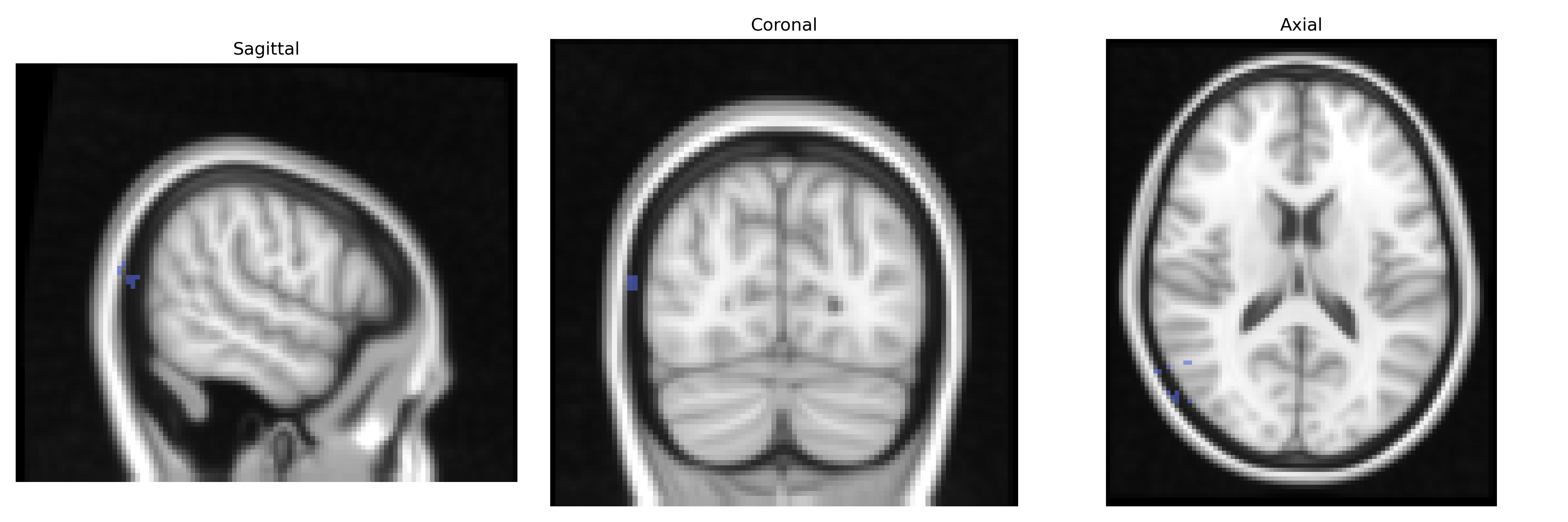
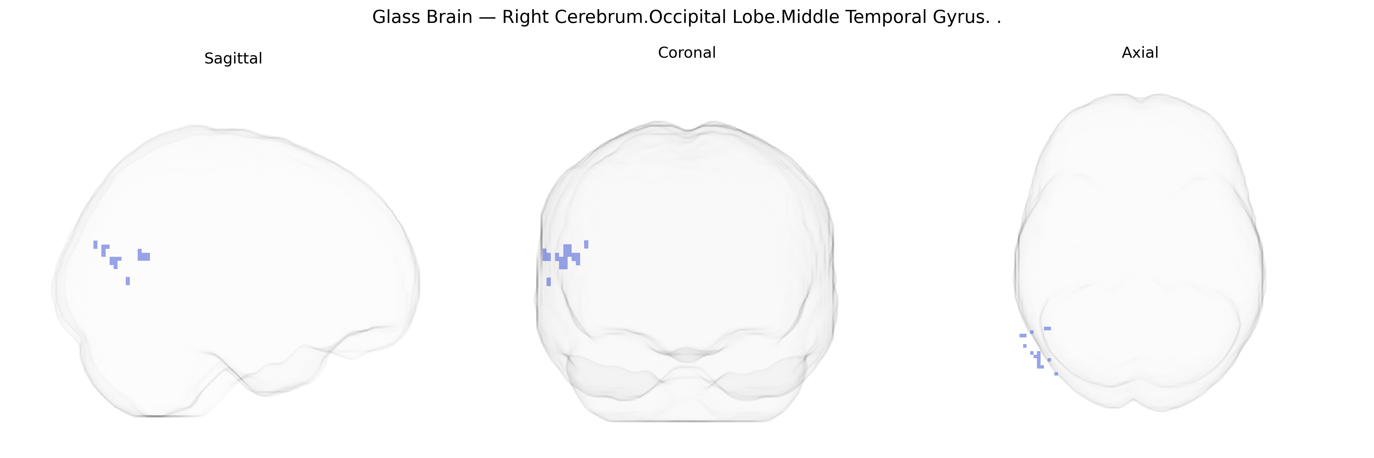
The bilateral Right Cerebrum.Occipital Lobe.Middle Temporal Gyrus corresponds to portions of the middle temporal gyrus extending into or adjacent to the occipital lobe on the right hemisphere, encompassing cortex involved in higher-order visual processing and multimodal integration. This region contributes to the analysis of complex visual stimuli, including motion, object features, and aspects of visual memory, and is functionally connected with both ventral (object-related) and dorsal (spatial/motion-related) visual pathways. It interfaces with temporal association cortices that support language comprehension, semantic processing, and audiovisual integration, reflecting its role as a convergence zone for visual information with other sensory and cognitive domains. There is no direct link for this composite Talairach label; a closely related structure is the Middle temporal gyrus.
The bilateral right middle temporal gyrus in the occipital lobe region (per Talairach 2 mm labels; more generally spanning lateral temporal–occipital association cortex) has been implicated in several genetic and GWAS-based neuroimaging findings, particularly through large consortia such as ENIGMA and UK Biobank. Cortical thickness and surface area measures in middle temporal cortex show significant SNP-based heritability and have been associated with variants in genes involved in neurodevelopment and synaptic function, including common variants near genes such as POU3F2, DACT1, and KIAA0586 in broader temporal-lobe imaging GWAS, although locus specificity to the exact Talairach-defined bilateral right region is typically limited by parcellation granularity. Structural and functional properties of the middle temporal gyrus have been linked to polygenic risk for schizophrenia, major depressive disorder, and autism spectrum disorder, with PRS studies showing associations between higher psychiatric polygenic load and altered cortical thickness, volume, or functional connectivity in temporal association areas that encompass or overlap this region. GWAS of reading ability, language-related traits, and educational attainment have identified distributed genetic influences on temporal–occipital language and semantic networks, with imaging-genetics work demonstrating that alleles influencing these traits often modulate activation and structure in middle temporal regions engaged in semantic processing, word recognition, and audiovisual integration. Additionally, rare variants and copy-number variants associated with neurodevelopmental disorders, including 22q11.2 deletion and other CNVs, frequently show downstream effects on temporal cortical morphology, and Alzheimer’s disease–related risk loci such as APOE ε4 and GWAS hits in CLU, PICALM, and BIN1 have been linked to patterns of cortical thinning and hypometabolism extending into lateral temporal regions, sometimes involving the middle temporal gyrus in later-stage disease. Overall, genetic studies support the middle temporal gyrus as a heritable association cortex node where common and rare variants influencing cognitive, language, and psychiatric phenotypes exert measurable effects on structure and function, though most findings are at the level of broader temporal-lobe parcels rather than the specific bilateral right Talairach-defined subregion.
Overview generated by GPT-4o (2026).
Region ID: 561
Hemisphere: bilateral
Atlas: Talairach labels 2mm
Full Quality Version: Download MP4
Full Quality Version: Download MP4
Wali Sidiqyar*, Gaurav Rudravaram*, Elyssa M. McMaster, Trent M. Schwartz, Adam M. Saunders, Kurt G. Schilling, Bennett A. Landman "Introducing SPINS: A Shared Public Visualization Library of Neuroanatomical Structures." Medical Imaging with Deep Learning- short paper
This resource is licensed under CC0 1.0 Universal (Public Domain).