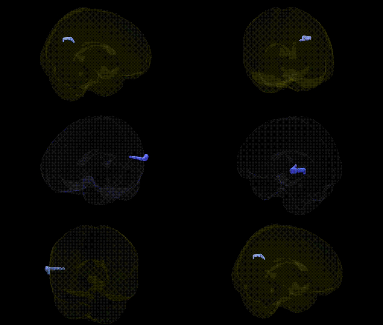
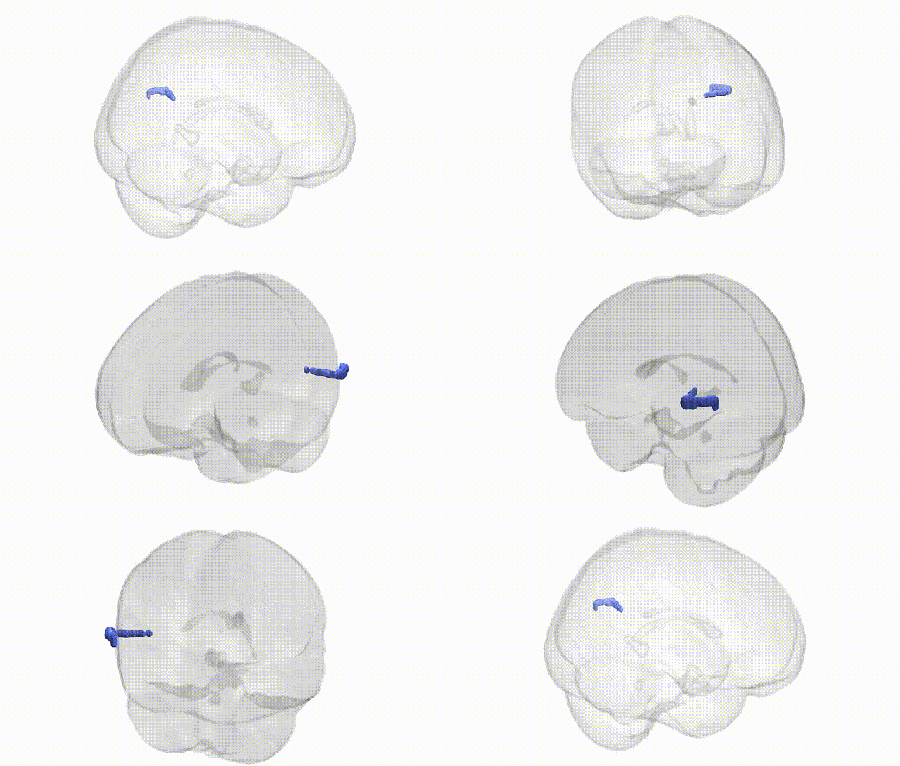
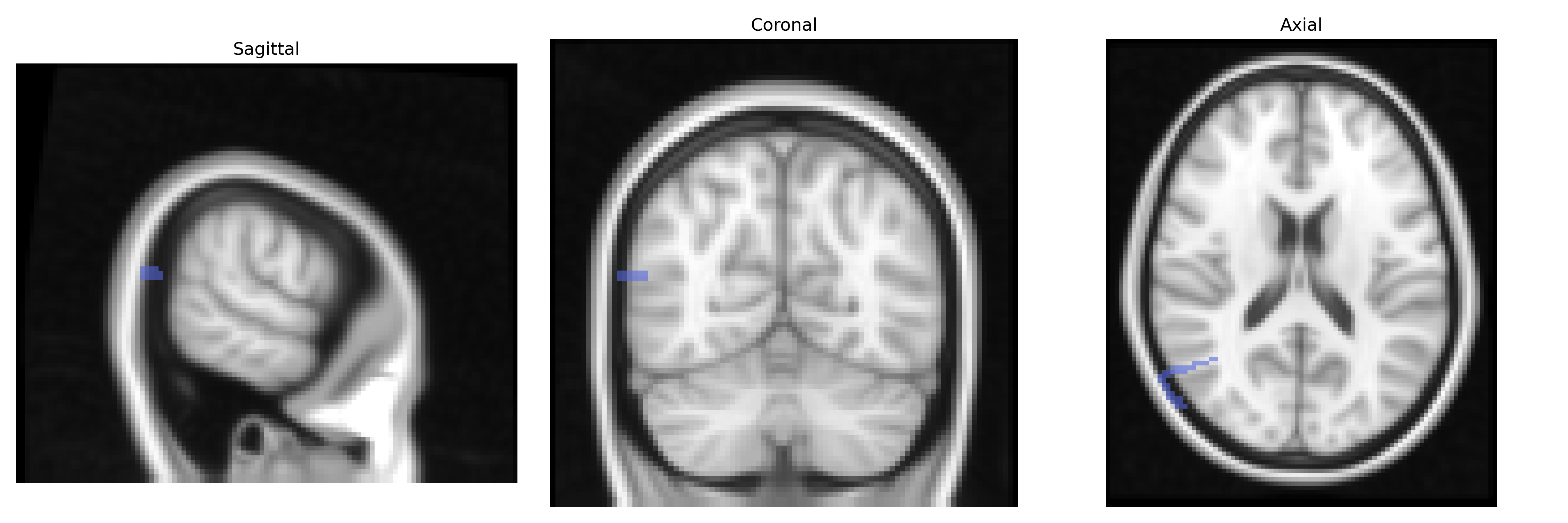
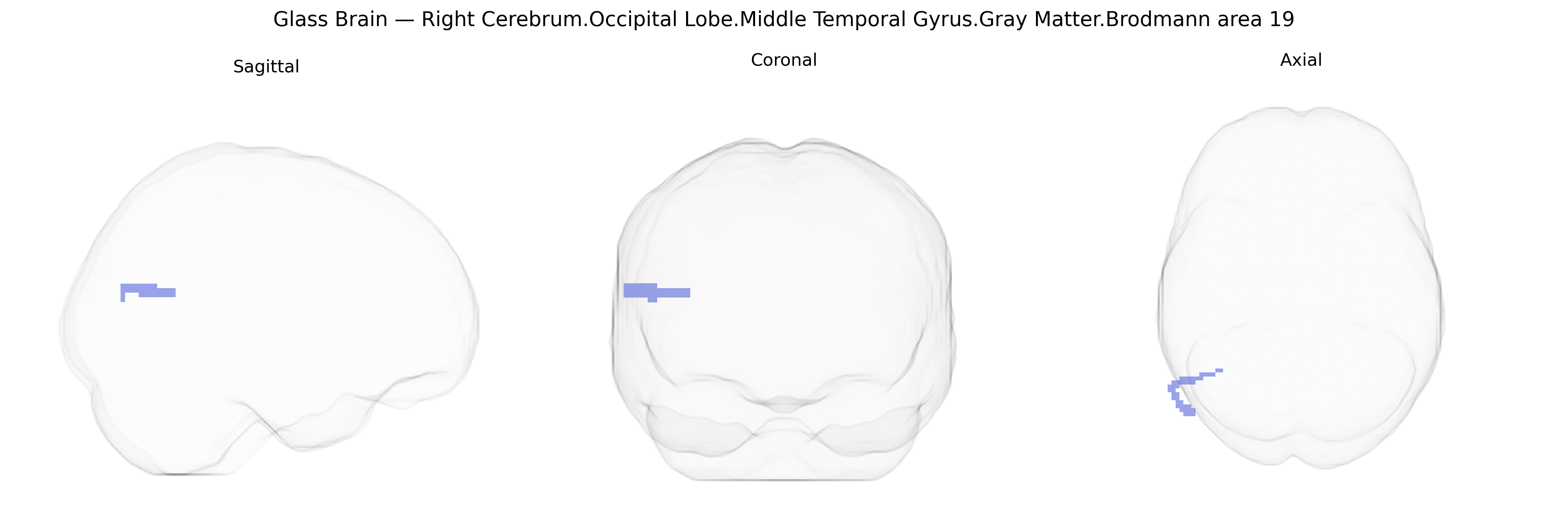
The bilateral Right Cerebrum Occipital Lobe Middle Temporal Gyrus Gray Matter Brodmann area 19 corresponds to a higher-order visual association region situated at the interface of the occipital and temporal cortices, involved in complex processing of visual information including object features, motion, and integration of visual stimuli with memory and semantic context. Although specifically labeled in the Talairach 2 mm Atlas as part of the middle temporal gyrus within the occipital lobe, functionally this territory contributes to the ventral and dorsal visual streams and participates in visual recognition, visuospatial analysis, and possibly multimodal integration with auditory and language-related areas in the temporal lobe. In neuroimaging and lesion studies, activity and damage in Brodmann area 19 are associated with impairments in visual perception and interpretation rather than primary visual loss, reflecting its role beyond basic sensory encoding. There is no direct Wikipedia article for this exact composite region; a closely related structure is Brodmann area 19.
The bilateral right middle temporal gyrus in the occipital lobe (Brodmann area 19; Talairach 2 mm atlas) lies within higher-order visual association cortex and has been implicated in genetic studies of visual processing, reading, and neuropsychiatric conditions. GWAS and imaging‑genetics work using cortical thickness, surface area, or voxel‑based morphometry have associated variants in genes such as MEF2C, POU3F2, KIAA0319, and DCDC2 with structural and functional differences in posterior temporal and occipito‑temporal regions related to dyslexia, reading ability, and language processing, which often extend into or near BA19. Polygenic risk for schizophrenia and bipolar disorder has been linked to altered gray matter and activation in posterior temporal–occipital regions, with risk alleles in genes including CACNA1C, GRIN2A, and complement pathway genes (e.g., C4), although these associations typically involve broader visual and language networks rather than BA19 in isolation. Autism‑spectrum GWAS and rare variant studies involving synaptic genes (e.g., SHANK3, NRXN1) have also been connected to atypical activation and connectivity in occipito‑temporal cortex during face and social perception tasks. Large ENIGMA‑style consortia have reported modest but significant SNP‑based heritability for cortical measures in the middle temporal and adjacent occipital association areas, suggesting that common genetic variation influences gray‑matter structure in this region, but specific, robust locus‑level associations unique to bilateral right BA19 remain limited, and most evidence comes from broader regional or network analyses rather than precise Talairach BA19 targets.
Overview generated by GPT-4o (2026).
Region ID: 700
Hemisphere: bilateral
Atlas: Talairach labels 2mm
Full Quality Version: Download MP4
Full Quality Version: Download MP4
Wali Sidiqyar*, Gaurav Rudravaram*, Elyssa M. McMaster, Trent M. Schwartz, Adam M. Saunders, Kurt G. Schilling, Bennett A. Landman "Introducing SPINS: A Shared Public Visualization Library of Neuroanatomical Structures." Medical Imaging with Deep Learning- short paper
This resource is licensed under CC0 1.0 Universal (Public Domain).