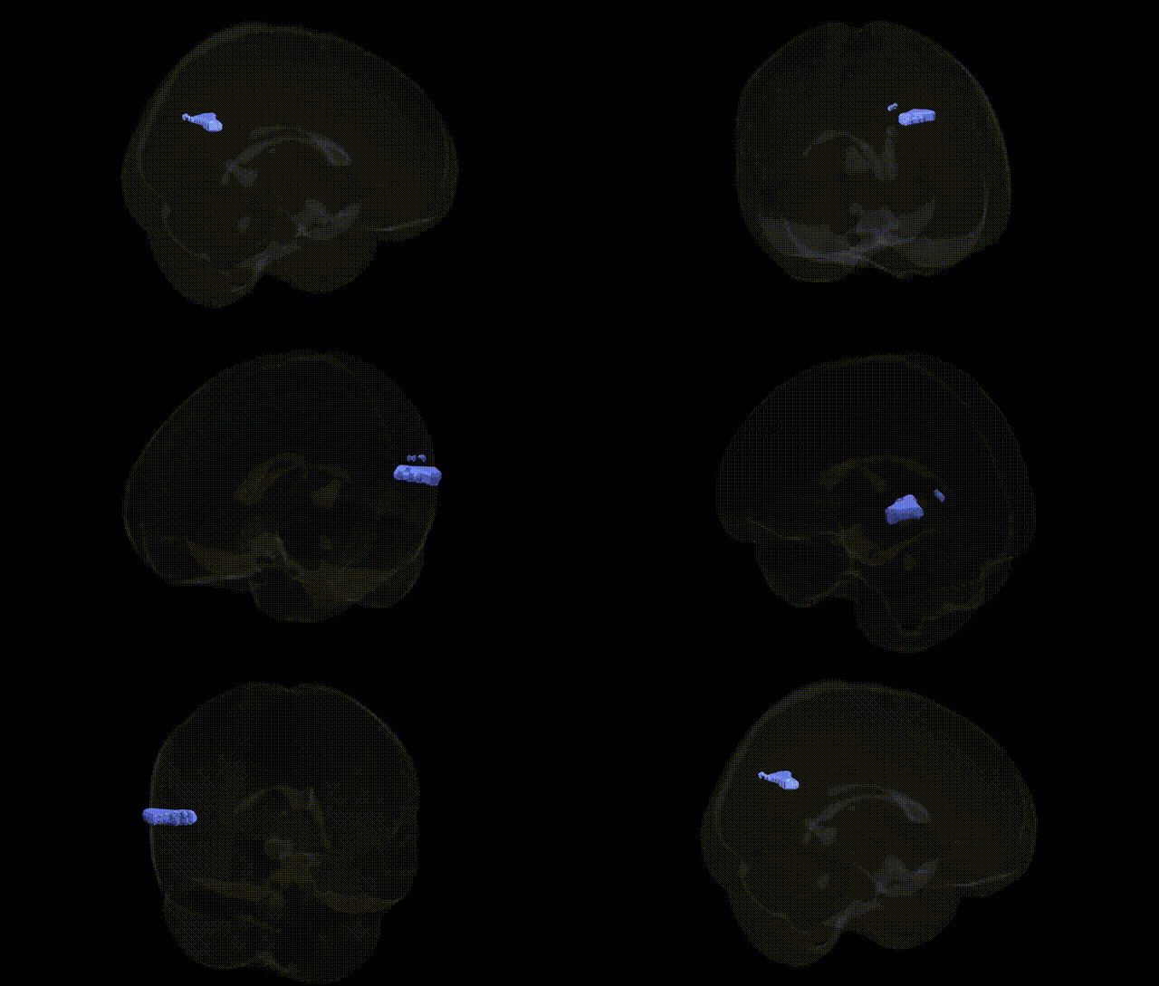
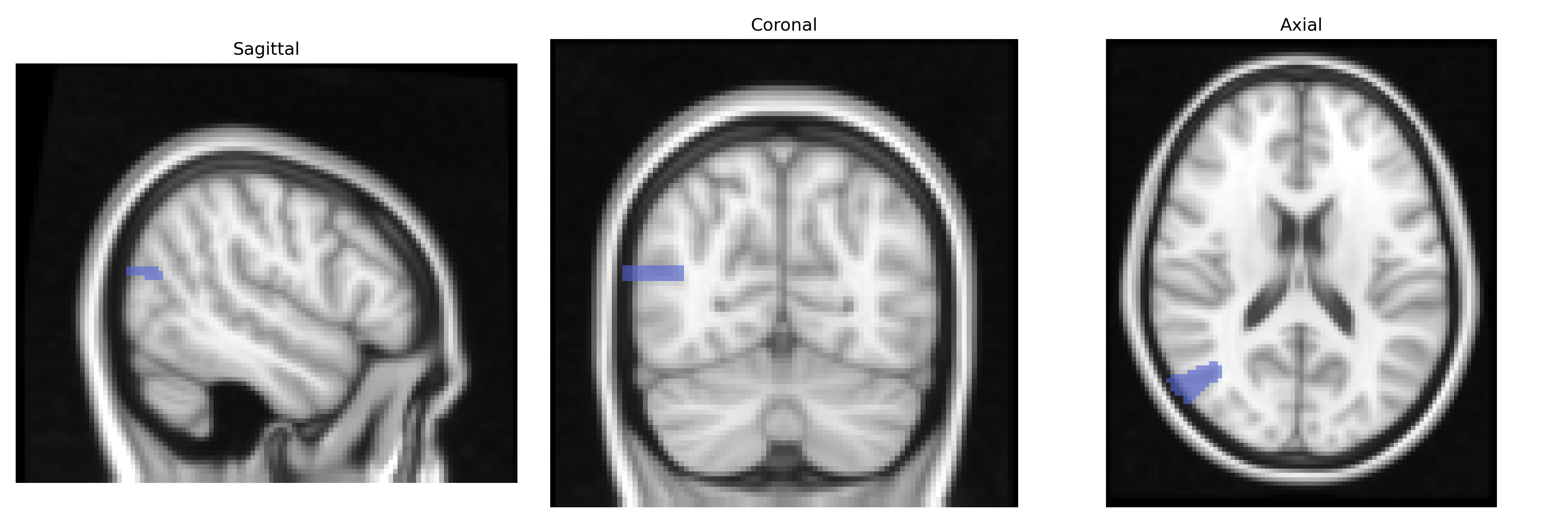
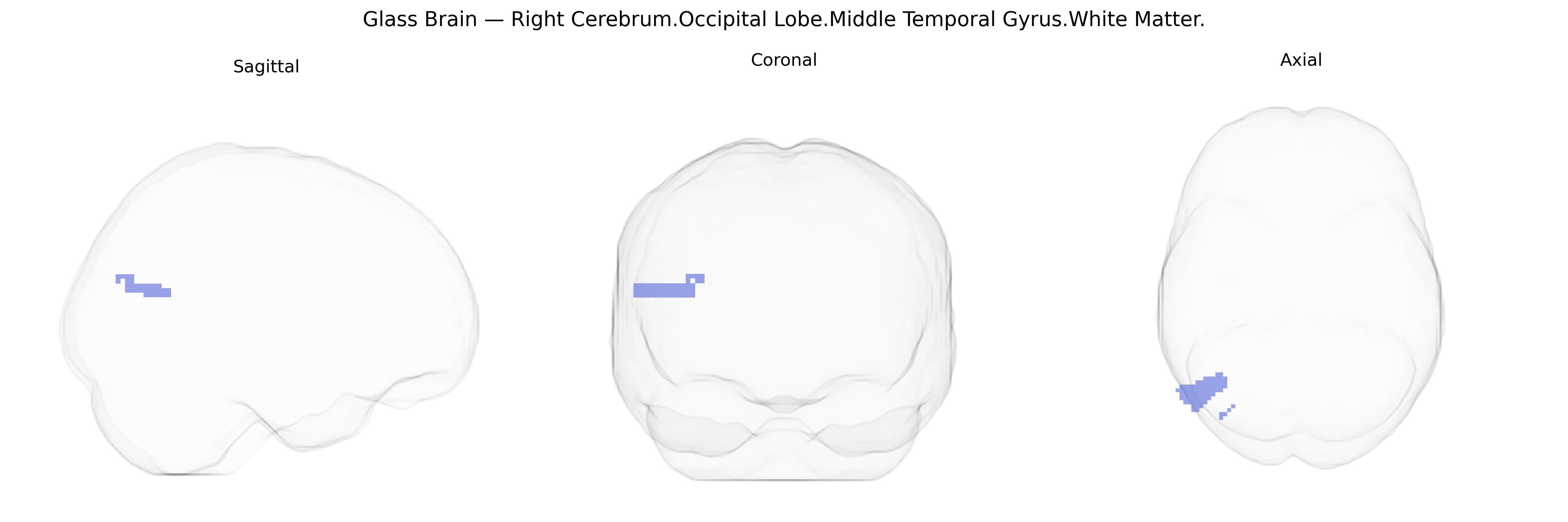
The bilateral Right Cerebrum.Occipital Lobe.Middle Temporal Gyrus.White Matter refers to the deep myelinated fiber tracts underlying the middle temporal gyrus in the right occipital–temporal junction region of the cerebral hemisphere. These white matter pathways support connectivity between the middle temporal gyrus—implicated in visual object recognition, semantic processing, and aspects of language—and adjacent occipital visual association cortices, as well as more distant cortical and subcortical structures via long-range tracts (e.g., portions of the inferior longitudinal fasciculus and inferior fronto-occipital fasciculus). Functionally, this white matter region contributes to the integration of high-order visual information with semantic and linguistic networks, facilitating the transmission of processed visual input toward temporal and frontal regions involved in memory and language. There is no direct Wikipedia article for this exact composite label; a related structure is the Middle temporal gyrus.
The bilateral Right Cerebrum Occipital Lobe Middle Temporal Gyrus White Matter, as defined in the Talairach 2 mm atlas, lies in a temporo-occipital association zone involved in visual processing, semantic integration, and higher-order perceptual functions, and genetic associations to this territory are typically inferred from imaging genetics and GWAS of MRI-based white-matter measures rather than from Talairach-specific studies. Large consortia such as ENIGMA and UK Biobank have identified variants in genes related to axonal guidance, myelination, and synaptic plasticity (e.g., CNTNAP2, NTRK1/2, PLEKHM1, and multiple loci in or near neurodevelopmental genes on chromosomes 3, 6, and 17) that are associated with fractional anisotropy and mean diffusivity in temporal and occipital white-matter tracts, including the inferior longitudinal fasciculus and adjacent fibers that traverse this region. GWAS and candidate-gene studies in schizophrenia, bipolar disorder, autism spectrum disorder, and major depressive disorder have linked risk variants—such as those in CACNA1C, ZNF804A, and loci within the extended MHC region—to structural and connectivity alterations in temporo-occipital white matter, with diffusion tensor imaging repeatedly showing microstructural abnormalities in fibers passing through the middle temporal and occipital lobes. In addition, polygenic risk scores for reading and language-related traits, educational attainment, and general cognitive ability show associations with white-matter integrity in posterior temporal–occipital regions, supporting a genetic contribution to variability in this area’s microstructure and its role in visual word form and language networks. Although few studies isolate the exact Talairach-defined “Middle Temporal Gyrus.White Matter” label, convergent evidence from GWAS of brain imaging, neuropsychiatric disorders, and cognitive traits indicates that genetic influences on neurodevelopmental and myelination pathways contribute to individual differences and disease-related alterations in the white matter of this temporo-occipital region.
Overview generated by GPT-4o (2026).
Region ID: 701
Hemisphere: bilateral
Atlas: Talairach labels 2mm
Full Quality Version: Download MP4
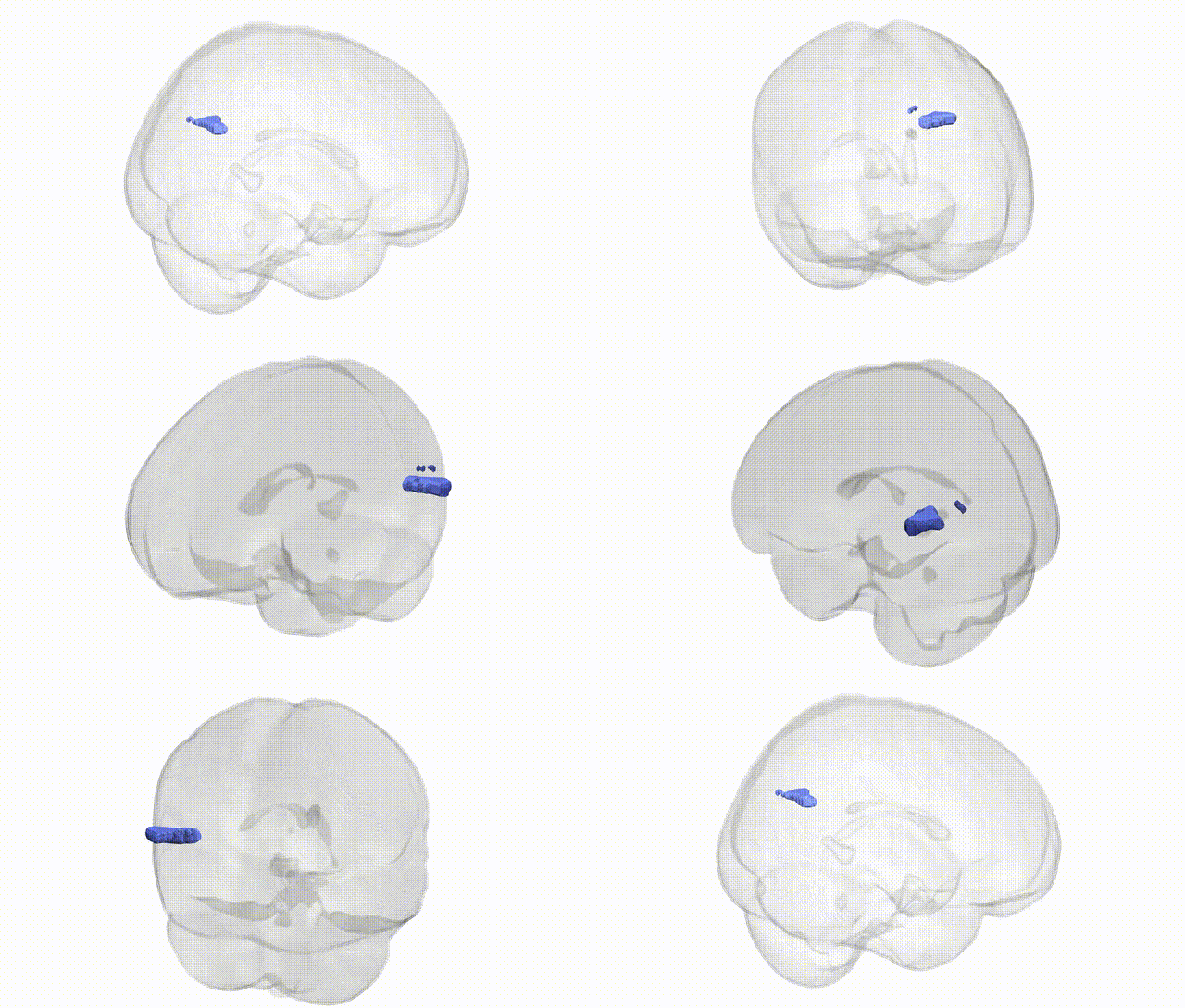
Full Quality Version: Download MP4
Wali Sidiqyar*, Gaurav Rudravaram*, Elyssa M. McMaster, Trent M. Schwartz, Adam M. Saunders, Kurt G. Schilling, Bennett A. Landman "Introducing SPINS: A Shared Public Visualization Library of Neuroanatomical Structures." Medical Imaging with Deep Learning- short paper
This resource is licensed under CC0 1.0 Universal (Public Domain).