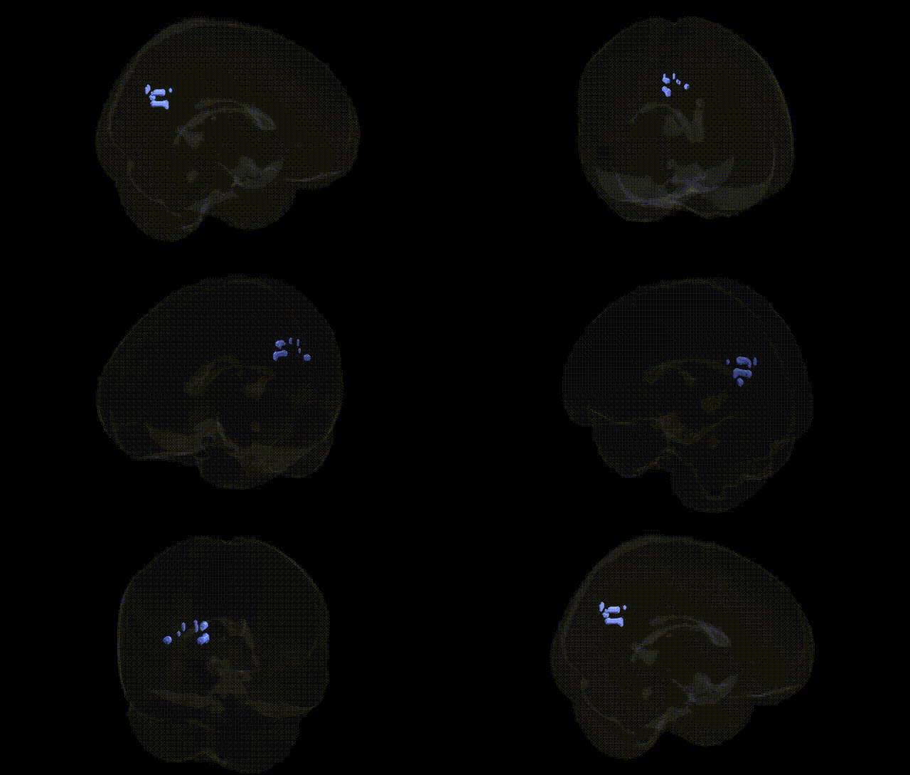
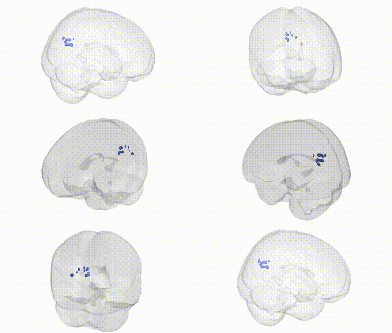
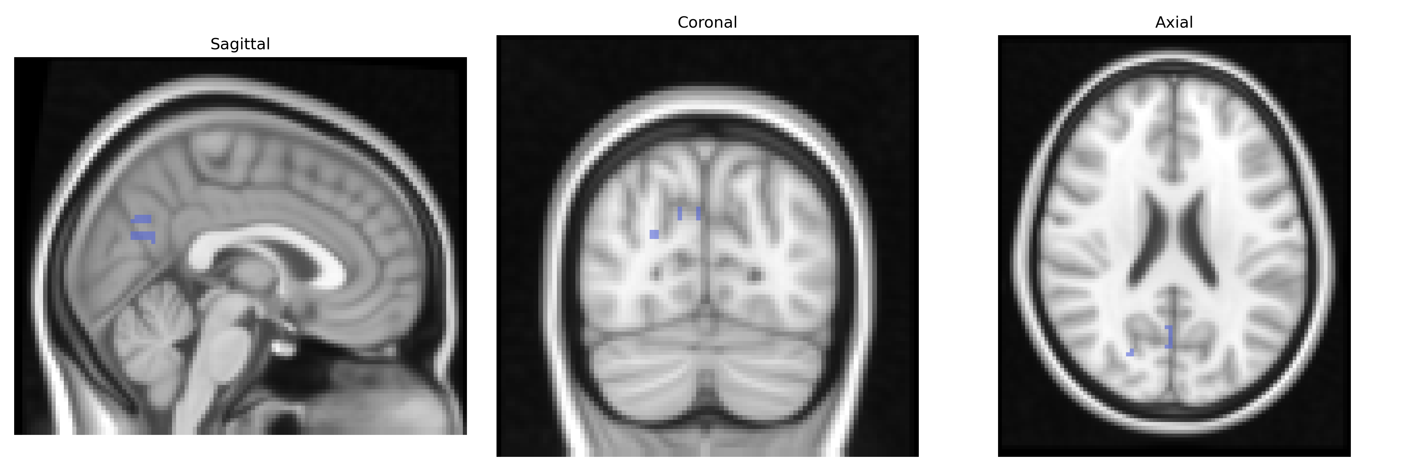
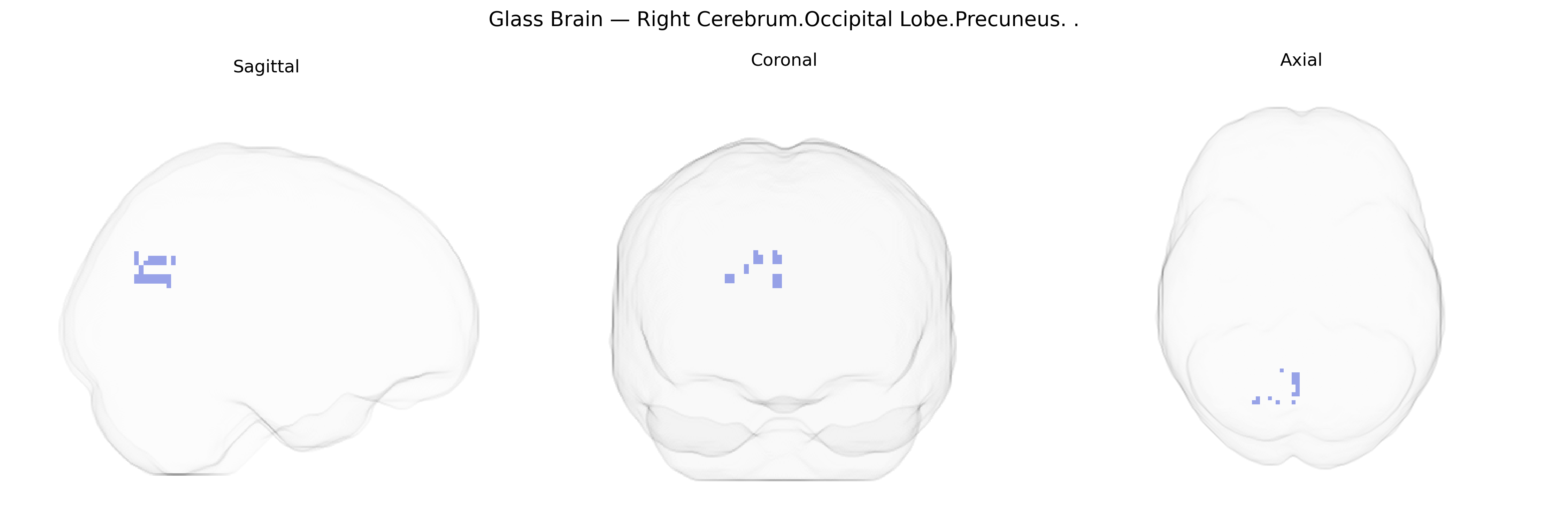
The bilateral Right Cerebrum.Occipital Lobe.Precuneus refers to the portion of the precuneus located within the right occipital lobe, as defined in the Talairach 2 mm Atlas. The precuneus is a medial parietal cortical region involved in visuospatial processing, self-related cognition, episodic memory retrieval, and aspects of consciousness, and it forms part of the default mode network. In this atlas labeling, the occipital-lobe-associated portion emphasizes its role in integrating visual information with higher-order associative processes, likely contributing to visual imagery, scene construction, and the transformation of visual inputs into more abstract representations. Functionally, this region participates in wide-ranging cortical networks, connecting with frontal, parietal, and limbic structures, and is implicated in tasks requiring mental imagery, perspective taking, and complex visual-spatial judgments. There is no direct link for this exact subregion; related information can be found under the broader cortical area Precuneus.
The bilateral right cerebrum occipital lobe precuneus (posterior medial parietal cortex bordering occipital regions in Talairach space) is a hub in the default mode network and has been repeatedly implicated in imaging genetics and GWAS of brain structure and function. Large-scale neuroimaging GWAS (e.g., ENIGMA, UK Biobank) have identified associations between precuneus/adjacent occipitoparietal cortical thickness and surface area with common variants in loci including genes involved in synaptic function and neurodevelopment such as BDNF, HMGA2, MEF2C, and regulatory regions near PDE4D and EPHB family members, although many signals are polygenic and spread across the genome. Resting-state connectivity of the precuneus shows heritability and has been linked to variants in genes related to glutamatergic signaling and calcium channels in imaging genetics of the default mode network. Disorders with robust precuneus involvement—such as Alzheimer’s disease, schizophrenia, major depressive disorder, ADHD, and autism spectrum disorder—have GWAS risk loci (e.g., APOE, CLU, CR1 for Alzheimer’s; CACNA1C, ZNF804A, GRM3 for schizophrenia; SLC6A4, BDNF, FKBP5 in stress-related depression; DRD4, DAT1/SLC6A3, FOXP1 and other neurodevelopmental genes for ADHD and ASD) that show convergent effects on precuneus structure, metabolism, or connectivity in case–control imaging studies. In Alzheimer’s disease, APOE ε4 carriers display early hypometabolism and atrophy in the precuneus/posterior cingulate region; in schizophrenia and bipolar disorder, polygenic risk scores correlate with altered precuneus activation during cognitive and self-referential tasks; and in depression and anxiety, risk variants in stress- and monoamine-related genes modulate precuneus involvement in rumination and default-mode hyperconnectivity. Additionally, GWAS of cognitive traits (general intelligence, episodic memory, and self-referential processing) have identified polygenic architectures that map onto precuneus volume and activation, suggesting that this region serves as a structural and functional mediator of genetically influenced higher-order cognition and vulnerability to neuropsychiatric disease, although specific single-gene–to–region mappings remain tentative and highly polygenic.
Overview generated by GPT-4o (2026).
Region ID: 828
Hemisphere: bilateral
Atlas: Talairach labels 2mm
Full Quality Version: Download MP4
Full Quality Version: Download MP4
Wali Sidiqyar*, Gaurav Rudravaram*, Elyssa M. McMaster, Trent M. Schwartz, Adam M. Saunders, Kurt G. Schilling, Bennett A. Landman "Introducing SPINS: A Shared Public Visualization Library of Neuroanatomical Structures." Medical Imaging with Deep Learning- short paper
This resource is licensed under CC0 1.0 Universal (Public Domain).