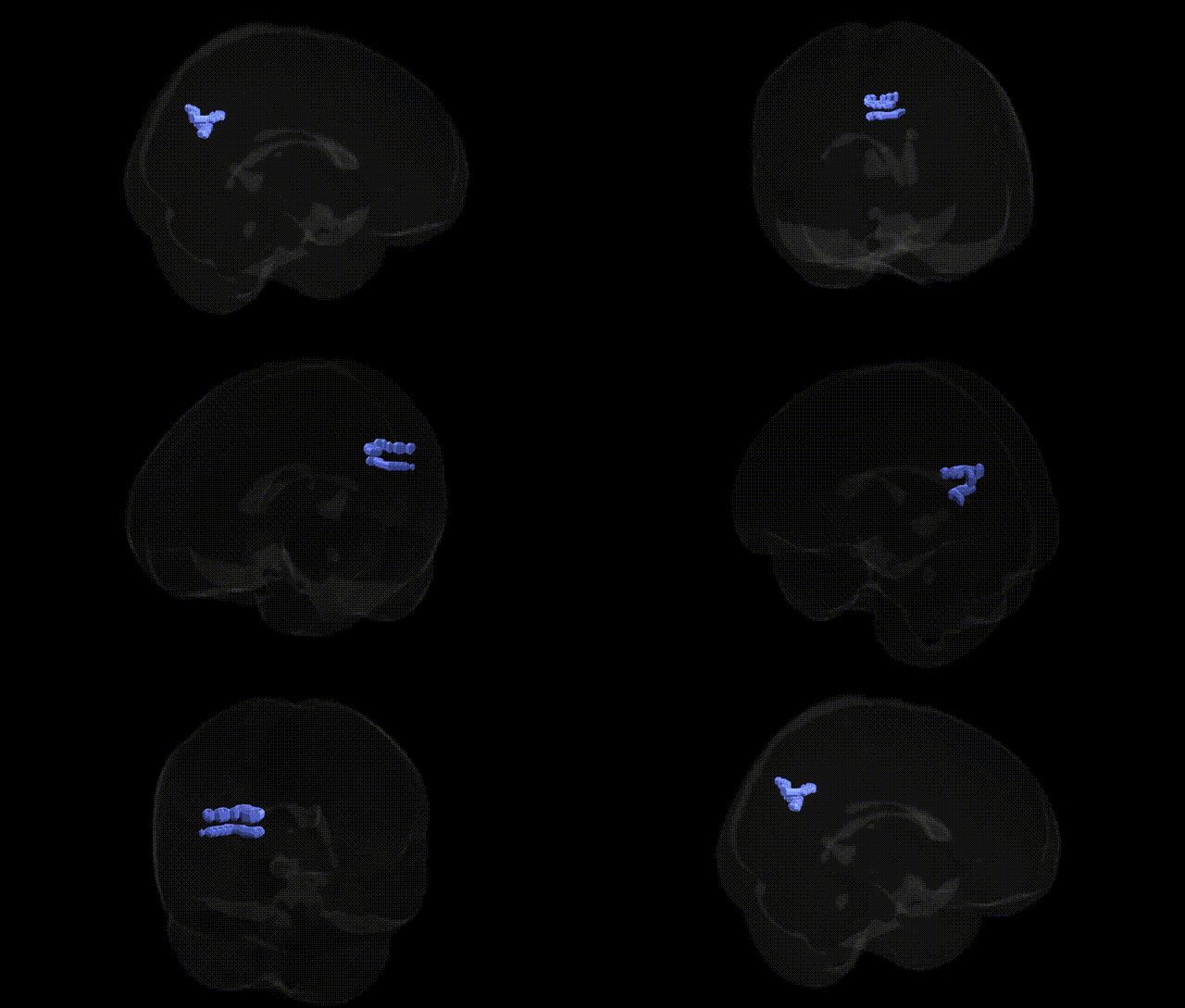
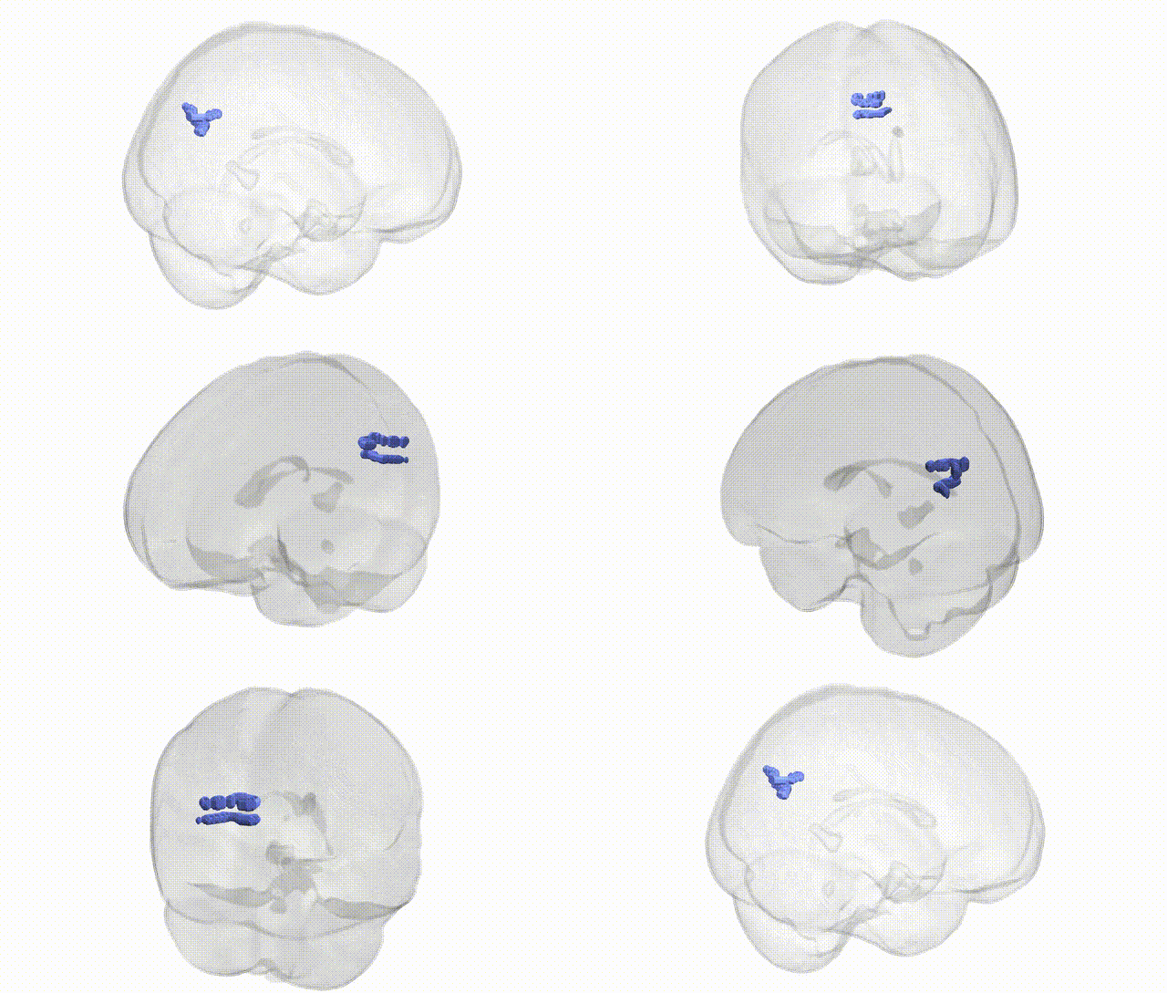
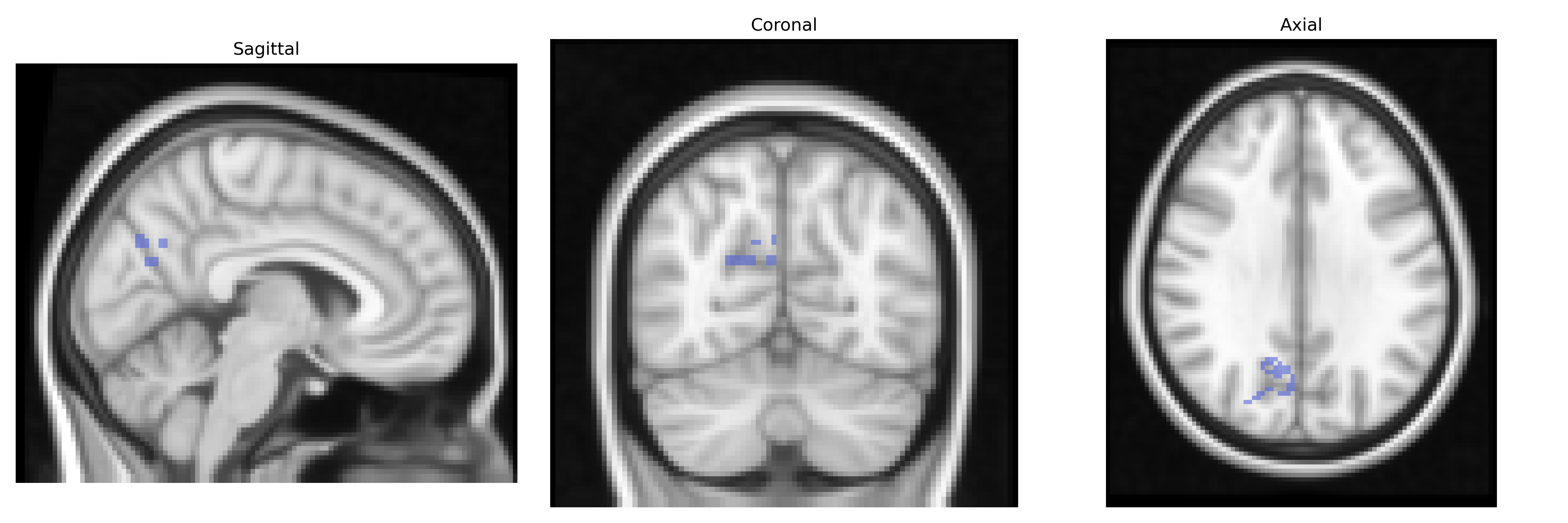
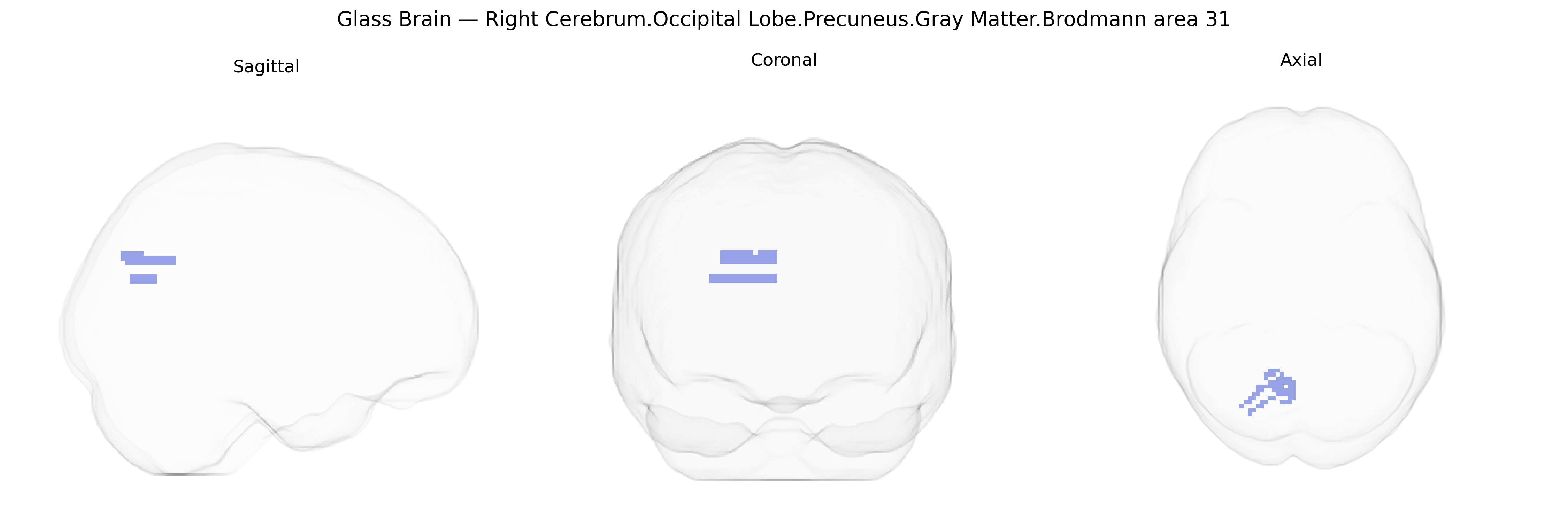
Bilateral Right Cerebrum Occipital Lobe Precuneus Gray Matter Brodmann area 31 corresponds to a posterior medial cortical territory situated near the parietal–occipital junction, forming part of the posterior cingulate/retrosplenial cortex and the medial aspect of the precuneus. Brodmann area 31 is cytoarchitectonically defined by its heteromodal association cortex, integrating visual, somatosensory, and limbic inputs, and is strongly implicated in episodic memory, self-referential and internally directed cognition, and navigation-related processing. Functionally, this region is a key node of the default mode network, demonstrating high metabolic activity at rest and deactivation during externally focused tasks, and it participates in large-scale networks subserving autobiographical memory retrieval, visuospatial imagery, and emotional appraisal. Structurally, it lies adjacent to other posterior cingulate and precuneal fields and exhibits dense connectivity with medial temporal structures, prefrontal association cortex, and other parietal regions. There is no direct Wikipedia article for “Brodmann area 31” as defined in the Talairach atlas; a closely related structure is the Posterior cingulate cortex.
The bilateral right precuneus in Brodmann area 31, a medial parietal/occipital region implicated in default mode network function, has been repeatedly highlighted in imaging genetics and GWAS of brain structure and connectivity, though specific studies rarely reference Talairach 2 mm labels explicitly. Large-scale neuroimaging GWAS (e.g., ENIGMA, UK Biobank) have identified common variants in genes related to synaptic development and plasticity (such as BDNF, CNTNAP2, and GRIN2B) and in neurodevelopmental regulatory loci that modulate cortical thickness and surface area in medial parietal and adjacent occipital cortices, which include the precuneus/BA31 territory. This region shows heritable variation in gray matter volume and resting-state connectivity, and its structure and function have been associated with genetic risk for Alzheimer’s disease (APOE and polygenic risk scores), schizophrenia (risk alleles in the MHC region and candidate synaptic genes), major depression, and autism spectrum disorder, often via altered default mode network connectivity. GWAS of cognitive traits and personality, particularly episodic memory, self-referential processing, and traits like openness and neuroticism, have also reported associations between polygenic scores for these traits and precuneus/BA31 morphology or activation patterns. Overall, genetic influences on this region appear to converge on pathways involved in neurodevelopment, synaptic signaling, and neurodegeneration, linking precuneus/BA31 structure and connectivity to risk for psychiatric and neurodegenerative disorders as well as individual differences in cognition and personality.
Overview generated by GPT-4o (2026).
Region ID: 824
Hemisphere: bilateral
Atlas: Talairach labels 2mm
Full Quality Version: Download MP4
Full Quality Version: Download MP4
Wali Sidiqyar*, Gaurav Rudravaram*, Elyssa M. McMaster, Trent M. Schwartz, Adam M. Saunders, Kurt G. Schilling, Bennett A. Landman "Introducing SPINS: A Shared Public Visualization Library of Neuroanatomical Structures." Medical Imaging with Deep Learning- short paper
This resource is licensed under CC0 1.0 Universal (Public Domain).