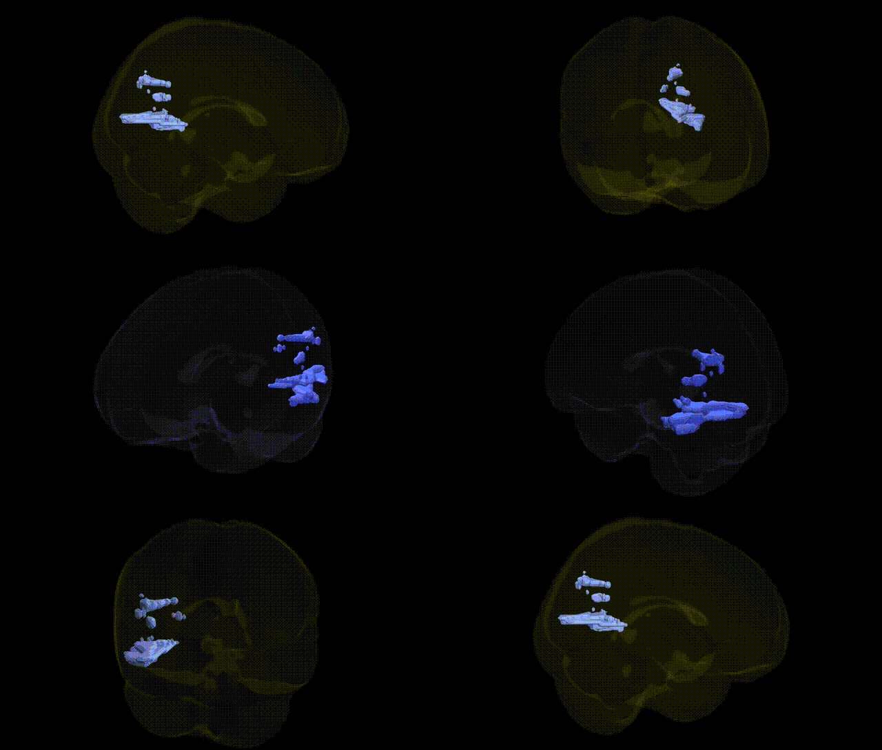
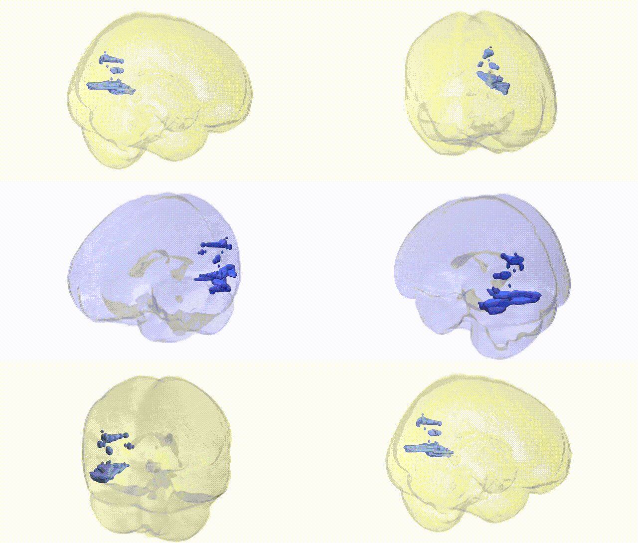
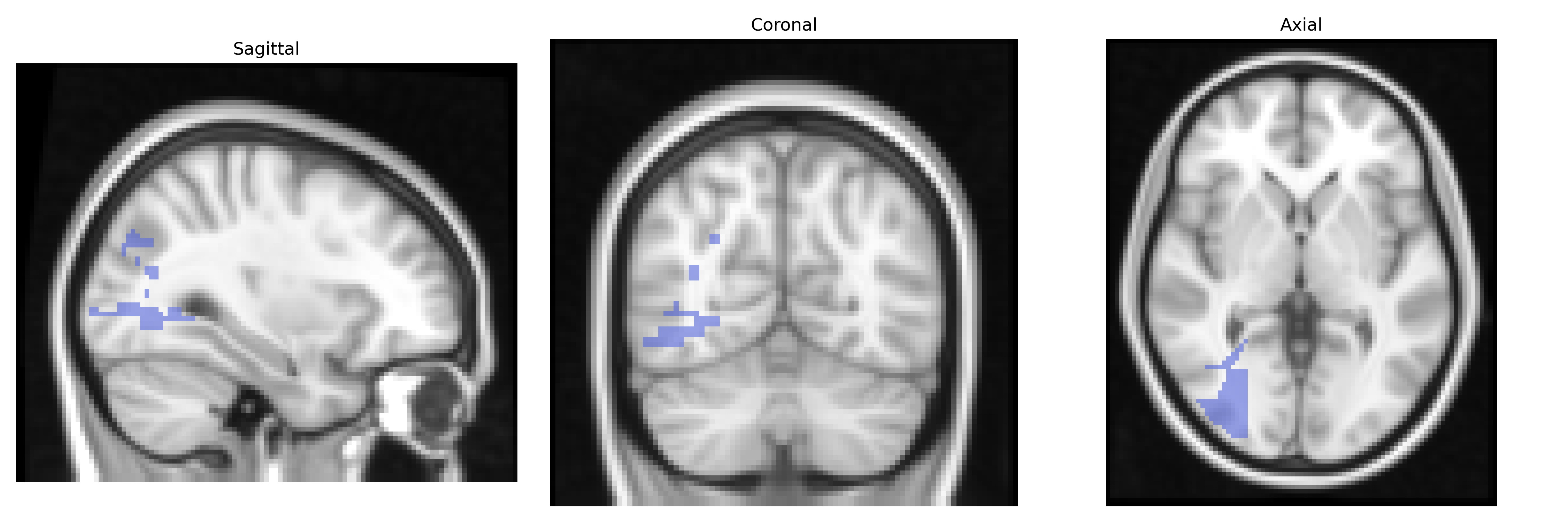
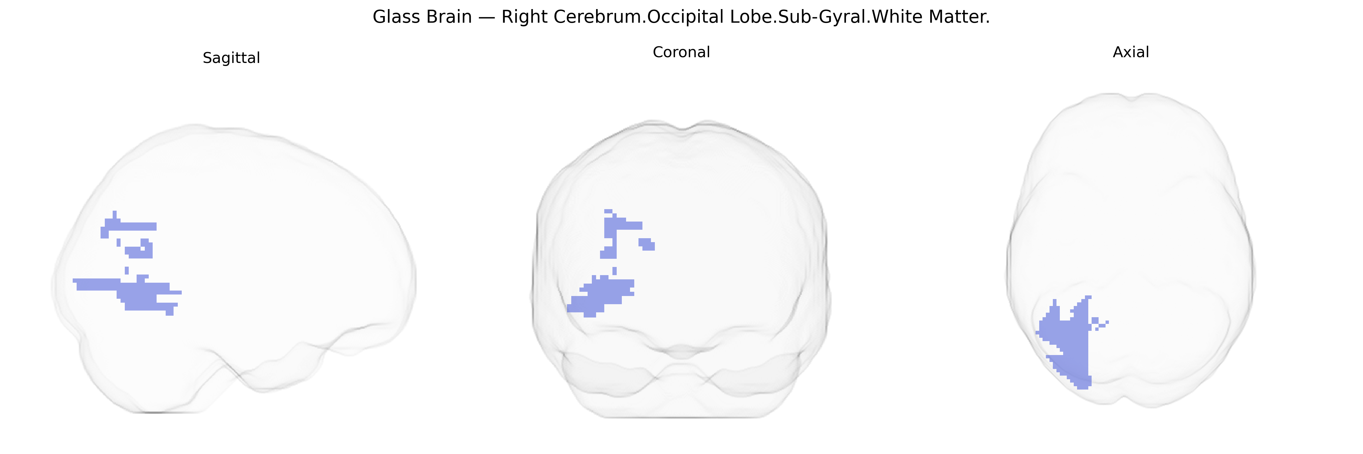
The bilateral Right Cerebrum.Occipital Lobe.Sub-Gyral.White Matter corresponds to deep white matter tracts located beneath the cortical gyri of the right occipital lobe, serving as conduits for visual information processing. These sub-gyral fibers include components of association, commissural, and projection pathways that link primary and secondary visual cortices with higher-order visual areas, parietal and temporal regions, and subcortical structures. Functionally, this white matter is essential for integrating visual input with spatial orientation, motion perception, and object recognition, contributing to coherent visual perception and visuospatial cognition. Damage to this region can disrupt visual field processing and visual integration without necessarily affecting the overlying cortical gray matter directly. There is no direct link for this specific sub-gyral white matter region; see the related Occipital lobe.
The bilateral Right Cerebrum Occipital Lobe Sub-Gyral White Matter region, as defined in the Talairach 2 mm atlas, corresponds largely to deep occipital white matter supporting visual processing networks, and genetic associations for this area arise mainly from imaging genetics and GWAS of brain structure rather than region-specific candidate studies. Large MRI-based GWAS (e.g., ENIGMA consortium and UK Biobank) have identified numerous loci—commonly involving genes such as MAPT, CNTNAP2, NRXN1, PDE4D, DCC, and variants near MEF2C and BDNF—that influence global and regional white matter microstructure (fractional anisotropy, mean diffusivity) and occipital or visual-tract integrity, including the optic radiations traversing occipital sub-gyral white matter. These genetic variants are frequently pleiotropic, showing overlap with risk loci for neurodevelopmental and psychiatric disorders (schizophrenia, autism spectrum disorder, ADHD, and major depression) and neurodegenerative conditions (Alzheimer’s disease, Parkinson’s disease), as well as cognitive and behavioral traits such as general intelligence, reading and visual processing skills, and susceptibility to hallucinations or visual perceptual disturbances, all of which rely on intact occipital white matter connectivity. However, due to the coarse spatial resolution and the emphasis on tract- or lobe-level measures in most GWAS, associations are typically reported for occipital white matter or visual pathways more broadly rather than specifically for the bilateral right sub-gyral occipital white matter parcel defined in the Talairach 2 mm atlas.
Overview generated by GPT-4o (2026).
Region ID: 320
Hemisphere: bilateral
Atlas: Talairach labels 2mm
Full Quality Version: Download MP4
Full Quality Version: Download MP4
Wali Sidiqyar*, Gaurav Rudravaram*, Elyssa M. McMaster, Trent M. Schwartz, Adam M. Saunders, Kurt G. Schilling, Bennett A. Landman "Introducing SPINS: A Shared Public Visualization Library of Neuroanatomical Structures." Medical Imaging with Deep Learning- short paper
This resource is licensed under CC0 1.0 Universal (Public Domain).