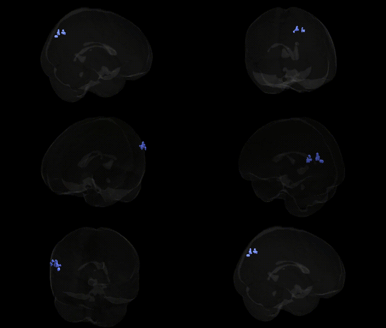
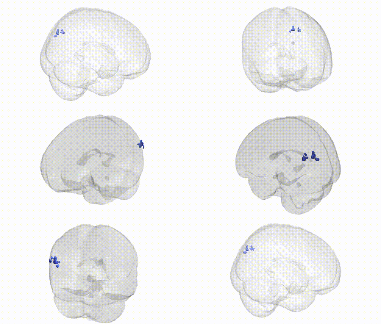
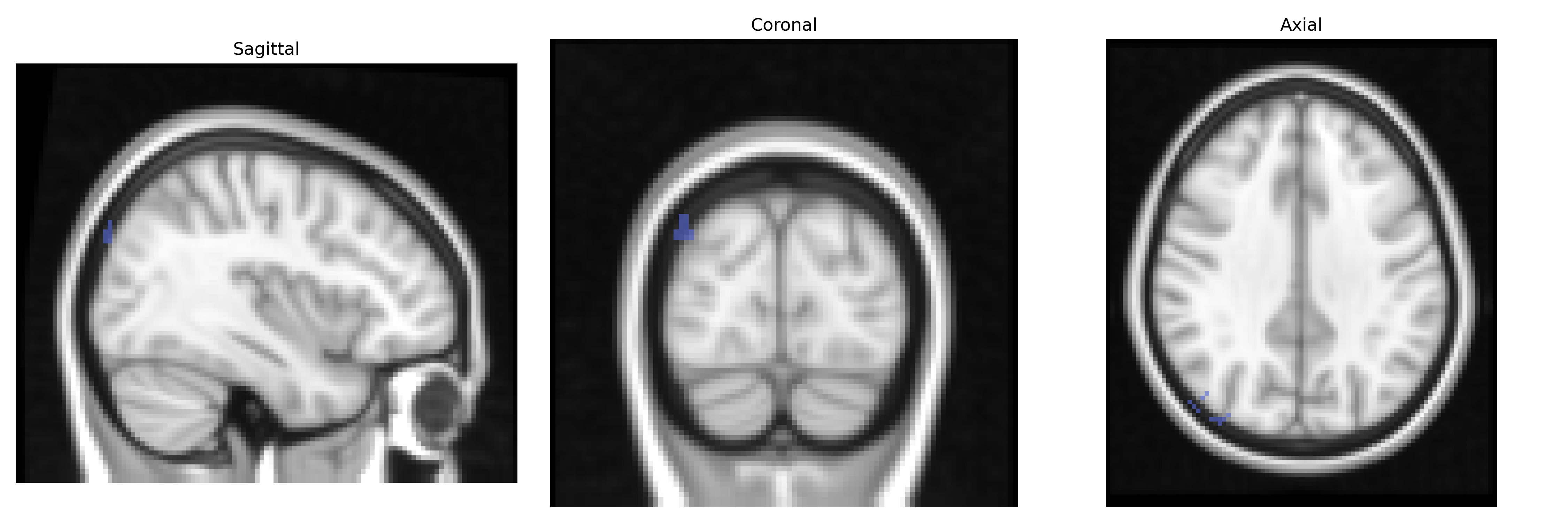
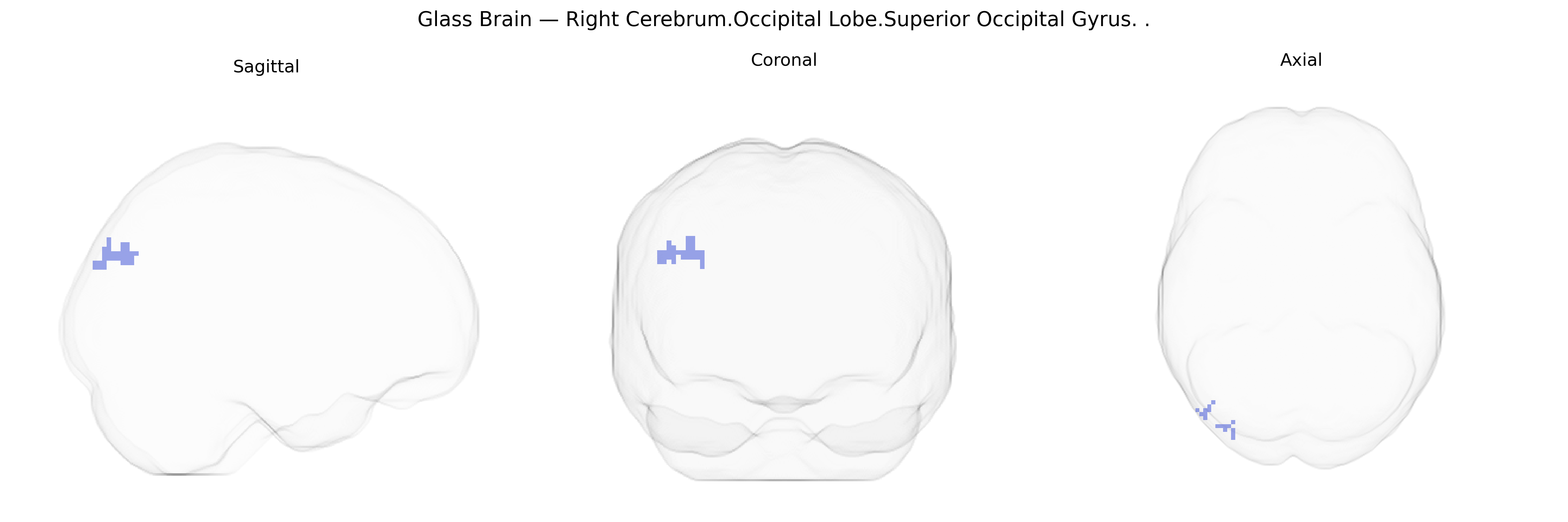
The bilateral Right Cerebrum.Occipital Lobe.Superior Occipital Gyrus, as defined in the Talairach 2 mm Atlas, refers to the dorsal portion of the occipital lobe on the right hemisphere, extending along the superior aspect of the lateral occipital surface and bordering parietal regions such as the precuneus and superior parietal lobule. This gyrus participates in higher-order visual processing, including aspects of spatial vision, motion perception, and visuospatial attention, and contributes to integration of visual information with parietal networks involved in visually guided behavior. Neuronal populations within this region are organized in a retinotopic manner, receiving input from earlier visual areas, and are interconnected with dorsal-stream pathways that support visually mediated actions and spatial cognition. There is no direct link for the superior occipital gyrus; a related structure is the Occipital lobe.
The bilateral right superior occipital gyrus (Right Cerebrum.Occipital Lobe.Superior Occipital Gyrus) is a higher-order visual association area implicated in genetic studies of brain structure, visual processing, and neuropsychiatric risk, although it is only rarely the primary focus of GWAS. Large imaging genetics consortia such as ENIGMA and UK Biobank have identified multiple loci (including variants near genes such as HMGA2, IGF1, and microtubule/axon guidance genes) associated with occipital cortical thickness and surface area, sometimes encompassing superior occipital territories, with polygenic influences overlapping those for general cognitive ability and educational attainment. Occipital cortex, including superior occipital regions, shows structural and functional alterations in disorders with known genetic components—such as schizophrenia, bipolar disorder, autism spectrum disorder, and major depression—and these changes often colocalize with GWAS-identified risk loci for synaptic function, neurodevelopment, and glutamatergic signaling, though attribution to this specific gyrus is typically indirect via parcellations. Genetic variants affecting myelination and visual pathway integrity (e.g., in MS susceptibility genes and genes related to white matter microstructure) have been associated with changes in occipital visual areas, and rare variants in neurodevelopmental genes can contribute to occipital cortical malformations and unusual visual processing phenotypes implicating superior occipital regions. Overall, current evidence supports a substantial but polygenic and distributed genetic contribution to the morphology and function of the superior occipital gyrus, with its associations largely embedded within broader occipital and visual system GWAS findings rather than region-specific loci.
Overview generated by GPT-4o (2026).
Region ID: 847
Hemisphere: bilateral
Atlas: Talairach labels 2mm
Full Quality Version: Download MP4
Full Quality Version: Download MP4
Wali Sidiqyar*, Gaurav Rudravaram*, Elyssa M. McMaster, Trent M. Schwartz, Adam M. Saunders, Kurt G. Schilling, Bennett A. Landman "Introducing SPINS: A Shared Public Visualization Library of Neuroanatomical Structures." Medical Imaging with Deep Learning- short paper
This resource is licensed under CC0 1.0 Universal (Public Domain).