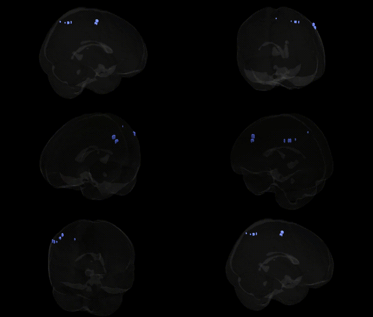
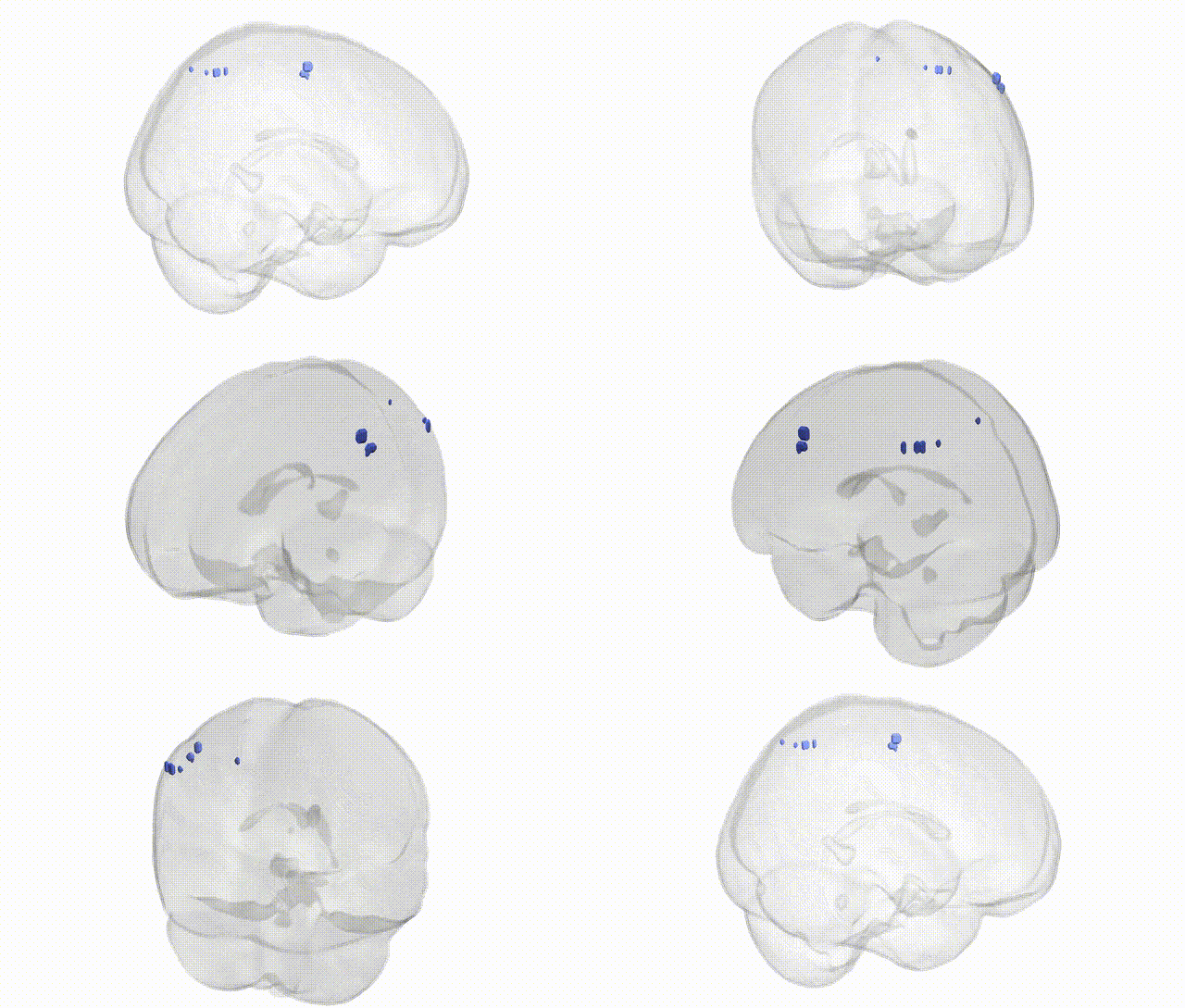
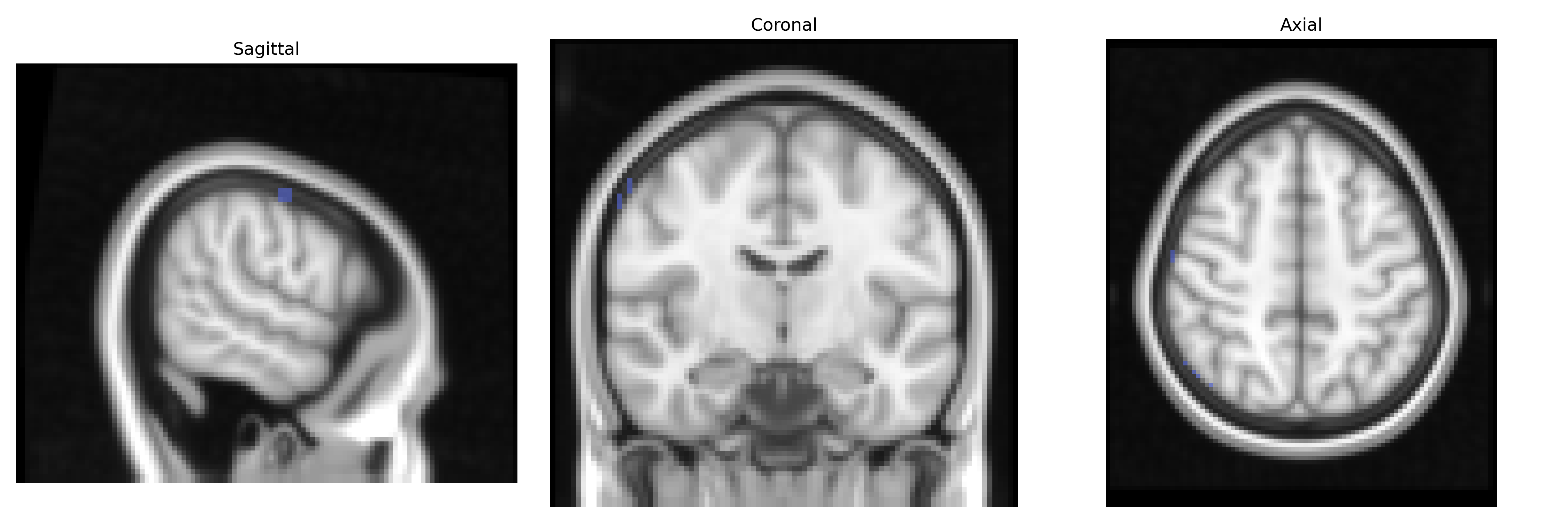
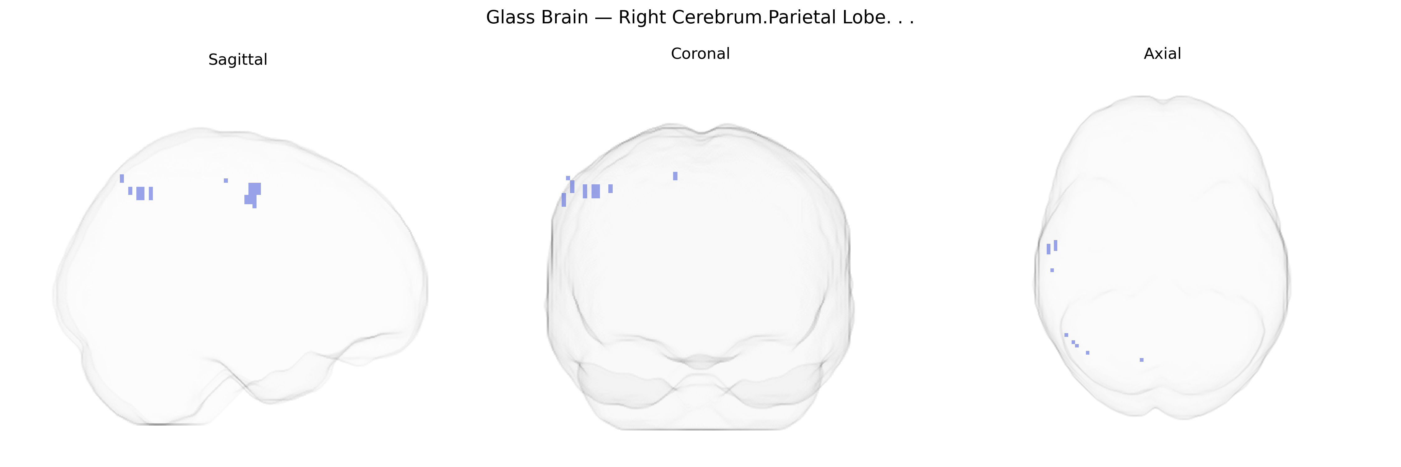
The bilateral Right Cerebrum.Parietal Lobe, as defined in the Talairach 2 mm atlas, encompasses parietal cortical regions of the right hemisphere that participate in multimodal sensory integration, spatial perception, and attentional control. This lobe receives and processes somatosensory input from the body, contributes to the representation of spatial relationships and body schema, and supports functions such as visuospatial cognition, mental rotation, and aspects of praxis. Functionally, right parietal areas are particularly implicated in hemispatial attention and awareness, and lesions are classically associated with deficits such as spatial neglect and impaired visuospatial processing. Anatomically, the parietal lobe is bounded anteriorly by the central sulcus, posteriorly by the parieto-occipital fissure, and superiorly and inferiorly by the longitudinal fissure and lateral sulcus, respectively, and contains key gyri including the postcentral gyrus, superior parietal lobule, and inferior parietal lobule.
Parietal lobe
The bilateral right parietal lobe (Talairach “Right Cerebrum.Parietal Lobe” region) has been repeatedly implicated in genetic studies of cognition, attention, and neuropsychiatric risk, although most findings derive from genome-wide association studies (GWAS) of imaging-derived phenotypes or clinical traits rather than Talairach labels specifically. Large neuroimaging GWAS consortia such as ENIGMA and UK Biobank have identified numerous common variants influencing parietal cortical thickness, surface area, and white-matter microstructure, with notable signals near genes involved in neurodevelopment and synaptic function, including variants in or near KIAA0586, TCF4, DLG2, GRIN2B, and CADM2. These genetically driven differences in parietal structure and connectivity have been associated with general cognitive ability, mathematical performance, visuospatial skills, and attentional control, consistent with the parietal lobe’s known functional roles. Parietal morphology and activation patterns also show heritable alterations in attention-deficit/hyperactivity disorder, autism spectrum disorder, schizophrenia, and major depressive disorder, with risk loci (e.g., in CACNA1C, MIR137, and complement pathway genes) linked to distributed cortical networks that include right parietal regions. Additionally, GWAS of stroke, Alzheimer’s disease, and cerebral small vessel disease report genetic effects on patterns of parietal atrophy and hypoperfusion, especially in posterior parietal areas, reflecting contributions from loci related to vascular biology (e.g., APOE, ACE) and amyloid processing. While these studies do not isolate the “bilateral Right Cerebrum.Parietal Lobe” Talairach label per se, they collectively support a strong genetic influence on structure and function of right parietal cortex and its involvement in the polygenic architecture of cognition, learning traits, and multiple psychiatric and neurological disorders.
Overview generated by GPT-4o (2026).
Region ID: 977
Hemisphere: bilateral
Atlas: Talairach labels 2mm
Full Quality Version: Download MP4
Full Quality Version: Download MP4
Wali Sidiqyar*, Gaurav Rudravaram*, Elyssa M. McMaster, Trent M. Schwartz, Adam M. Saunders, Kurt G. Schilling, Bennett A. Landman "Introducing SPINS: A Shared Public Visualization Library of Neuroanatomical Structures." Medical Imaging with Deep Learning- short paper
This resource is licensed under CC0 1.0 Universal (Public Domain).