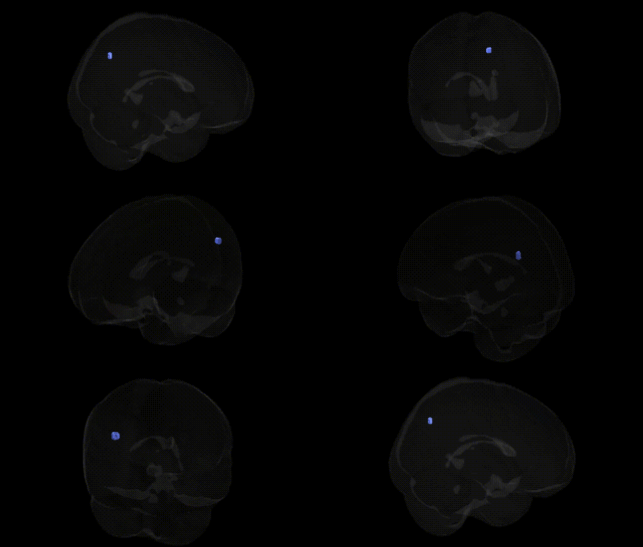
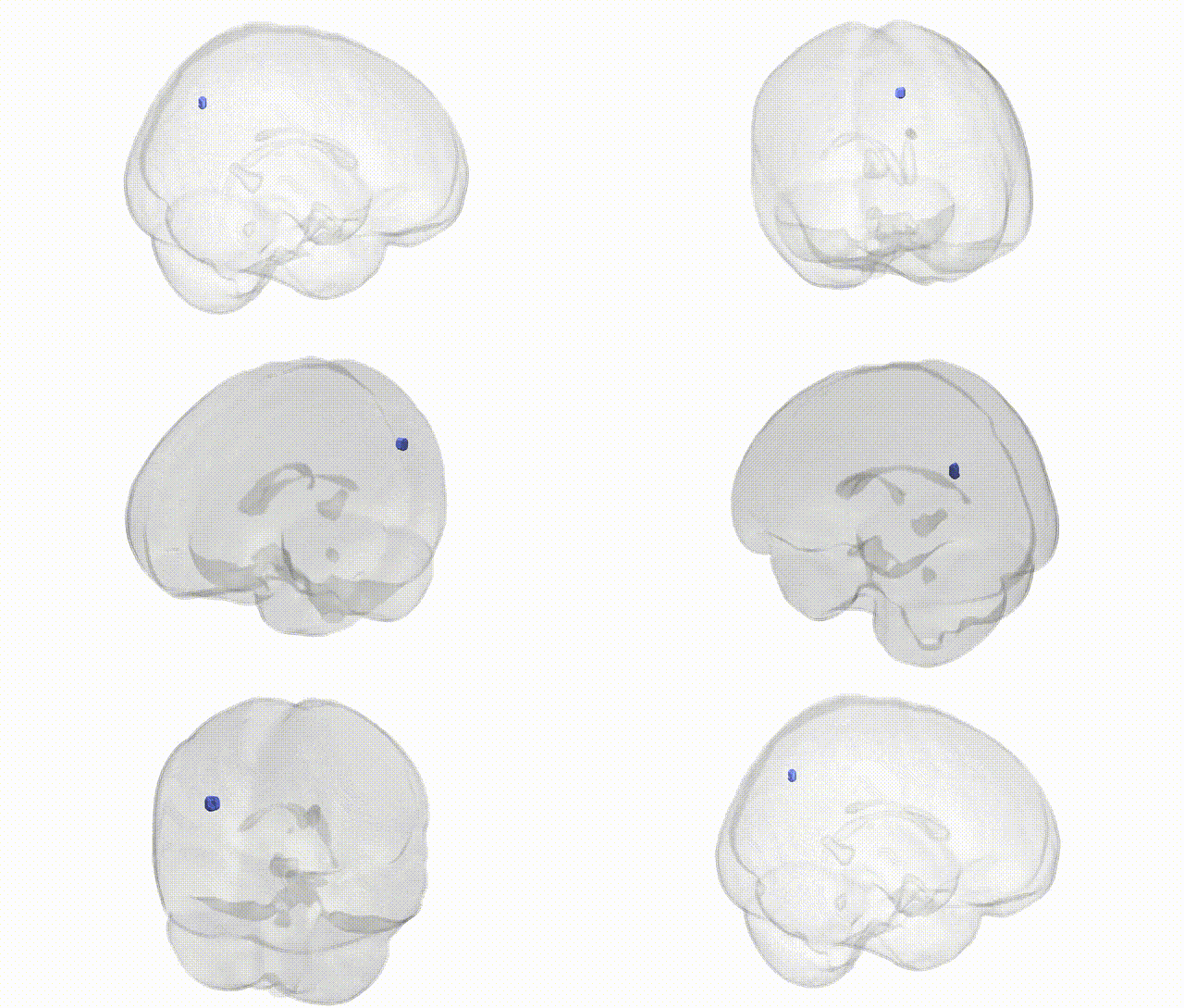
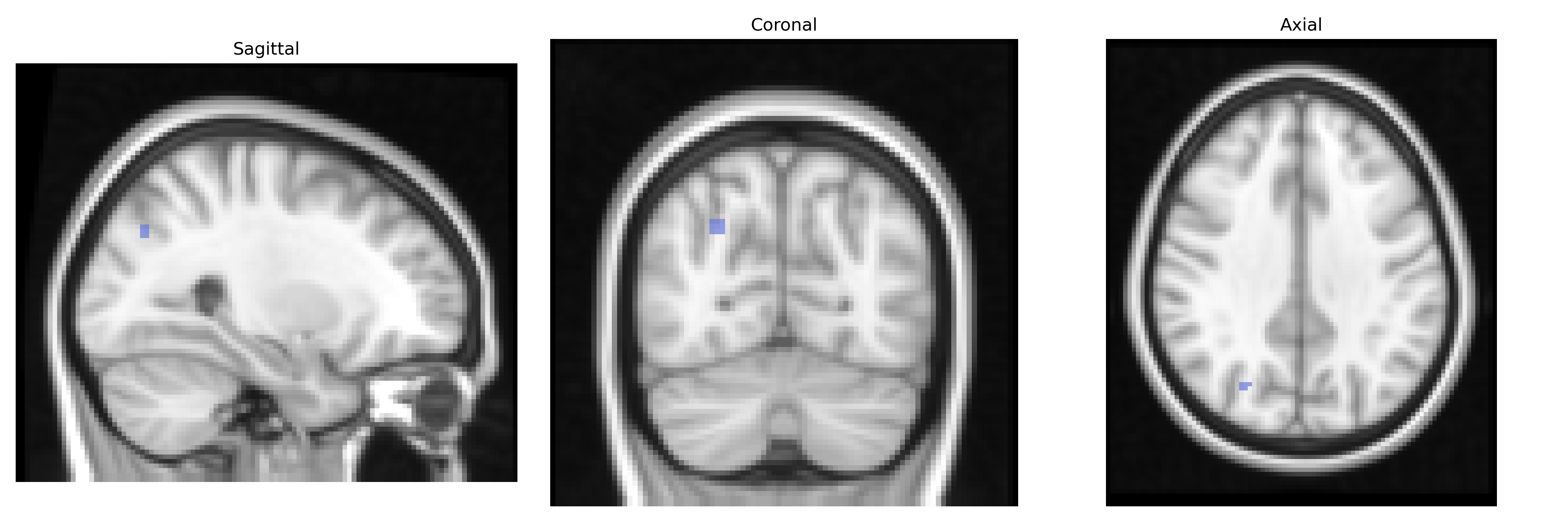
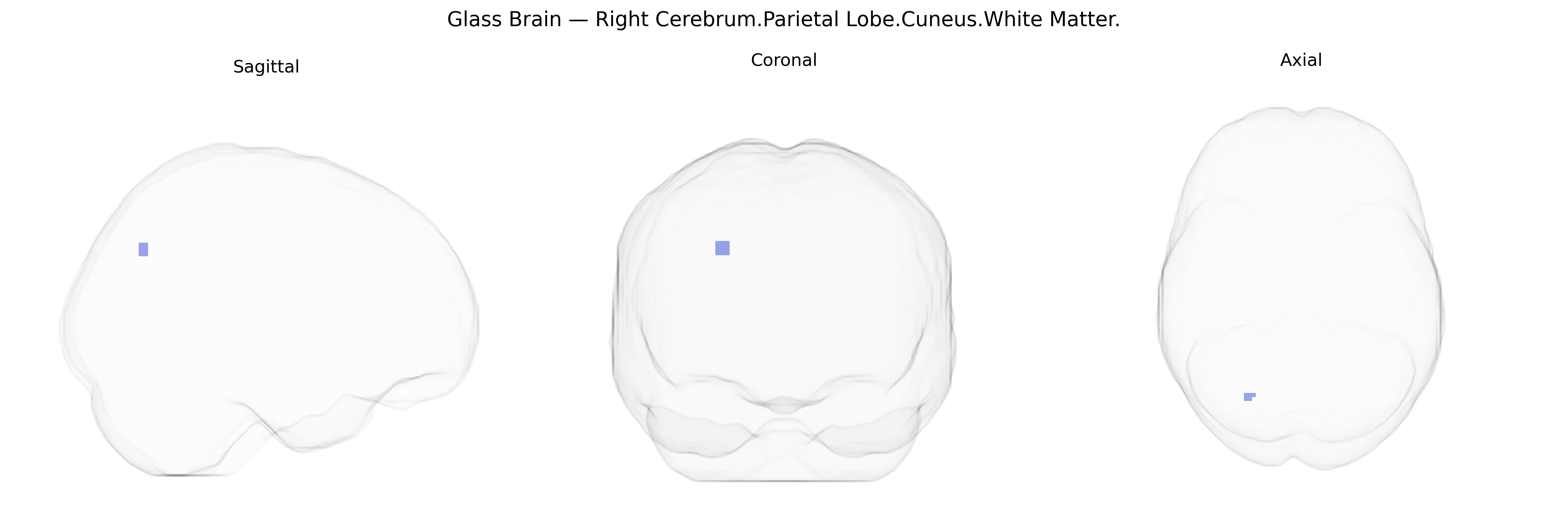
The bilateral Right Cerebrum.Parietal Lobe.Cuneus.White Matter corresponds to the subcortical fiber tracts underlying the cuneus region of the parietal lobe, situated on the medial surface of the occipital-parietal junction, dorsal to the calcarine sulcus. These white matter pathways contain association and projection fibers that interconnect the cuneus cortex with other visual, parietal, and higher-order associative areas, supporting integration and transmission of visual-spatial and visuomotor information. Although the Talairach label specifies parietal lobe, the cuneus is classically regarded as part of the medial occipital cortex involved in basic and higher-order visual processing. There is no direct link for this specific white matter subdivision; a closely related cortical region is the Cuneus.
Genetic associations specifically targeting the bilateral Right Cerebrum.Parietal Lobe.Cuneus.White Matter as defined in the Talairach 2 mm atlas are rarely reported at that fine-grained level, but convergent evidence from GWAS and imaging‑genetics studies implicates the cuneus and adjacent parietal-occipital white matter in several heritable traits and disorders. Large-scale MRI GWAS (e.g., ENIGMA, UK Biobank) show that regional cortical thickness, surface area, and white‑matter microstructure (fractional anisotropy, mean diffusivity) in posterior parietal/occipital regions, including the cuneus, are substantially heritable and influenced by common variants in genes related to axonal guidance, myelination, and synaptic development (such as loci near MAPT, WNT signaling genes, and neurodevelopmental transcription factors). Variants associated with general cognitive ability, educational attainment, and visual-spatial processing have been linked to structural measures in this territory, while polygenic risk for schizophrenia, bipolar disorder, major depressive disorder, and ADHD shows replicated associations with altered cuneus and parietal white‑matter integrity and cortical morphology, suggesting that genetic liability to these disorders partly manifests in this network. In Alzheimer’s disease and other dementias, GWAS loci such as APOE, BIN1, and CLU are associated with accelerated atrophy and disrupted white‑matter connectivity in posterior cortical regions that encompass the cuneus, consistent with the role of these genes in amyloid processing, lipid transport, and neuroinflammation. Additionally, individual GWAS of brain connectivity and resting-state networks highlight genetically influenced variation in posterior default mode and visual networks crossing the cuneus and parietal lobe white matter, linking this region to traits such as mind-wandering, visuospatial attention, and susceptibility to migraine and visual aura, although most findings remain at the level of broader occipital-parietal territories rather than the exact Talairach-defined parcel.
Overview generated by GPT-4o (2026).
Region ID: 964
Hemisphere: bilateral
Atlas: Talairach labels 2mm
Full Quality Version: Download MP4
Full Quality Version: Download MP4
Wali Sidiqyar*, Gaurav Rudravaram*, Elyssa M. McMaster, Trent M. Schwartz, Adam M. Saunders, Kurt G. Schilling, Bennett A. Landman "Introducing SPINS: A Shared Public Visualization Library of Neuroanatomical Structures." Medical Imaging with Deep Learning- short paper
This resource is licensed under CC0 1.0 Universal (Public Domain).