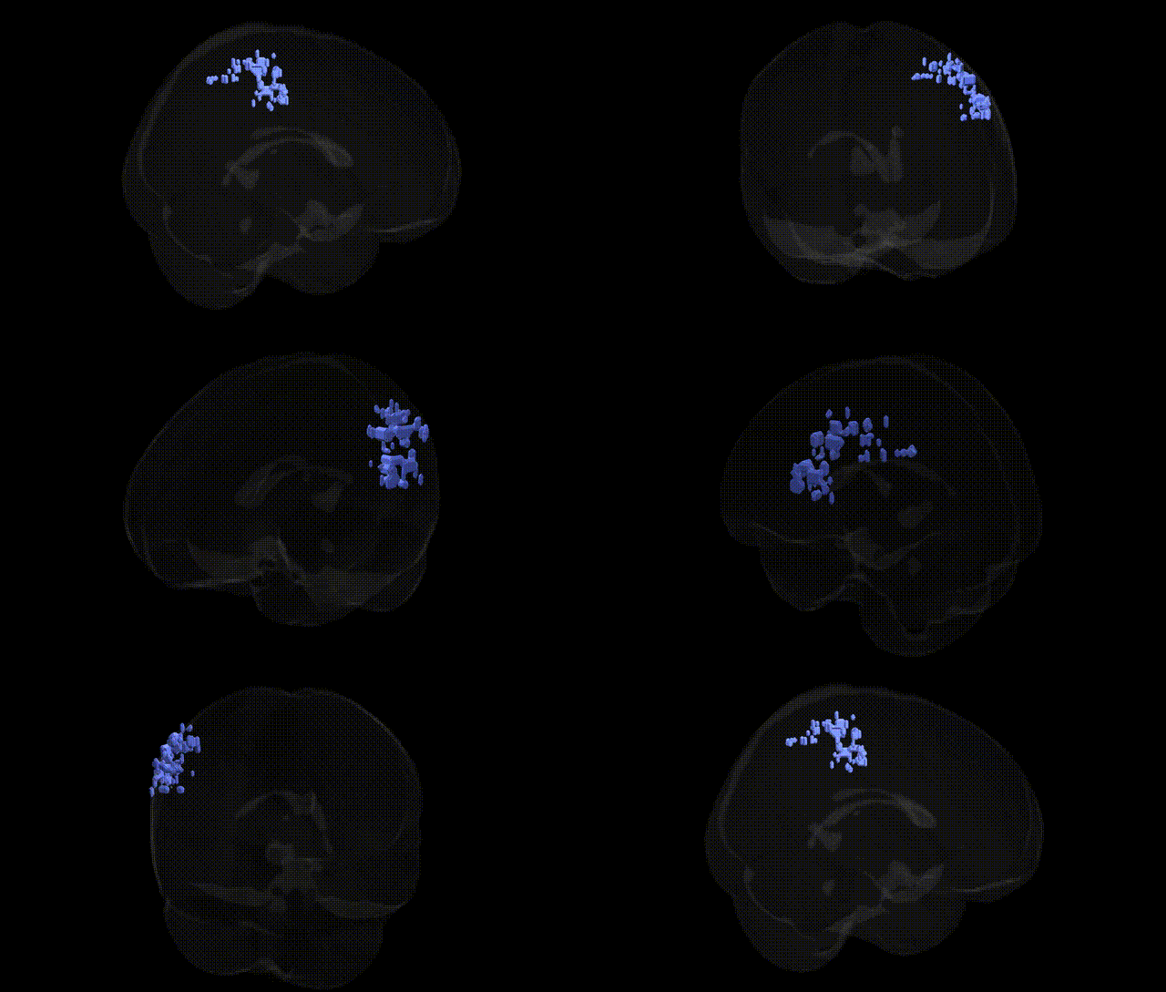
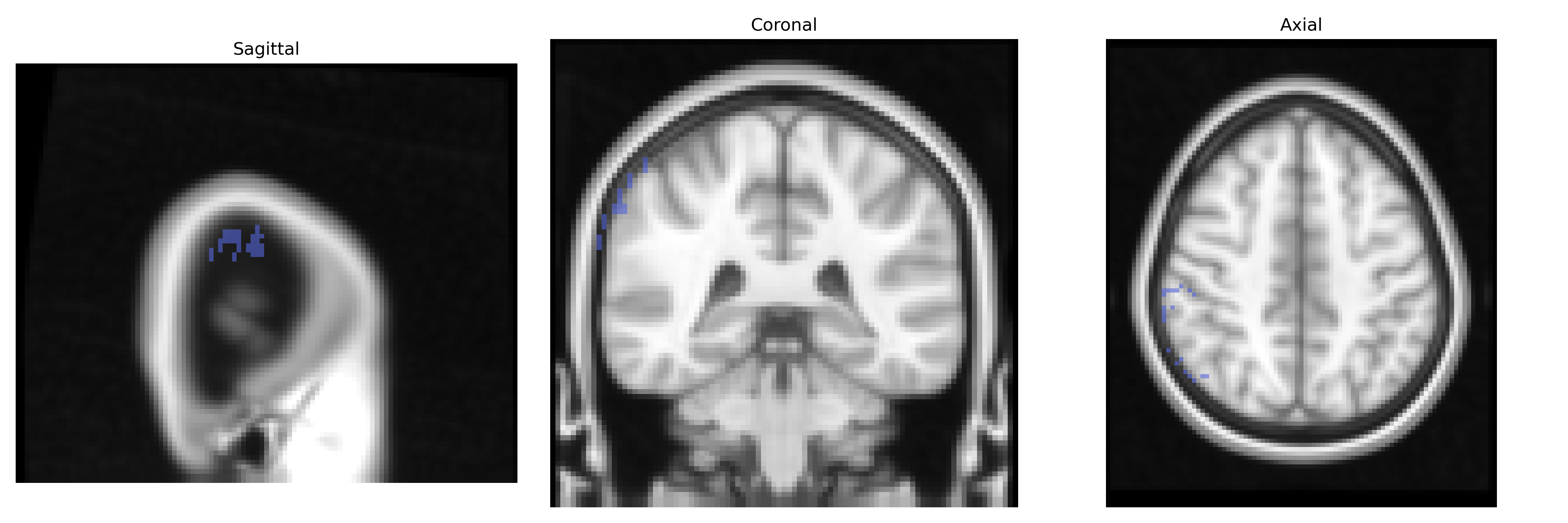
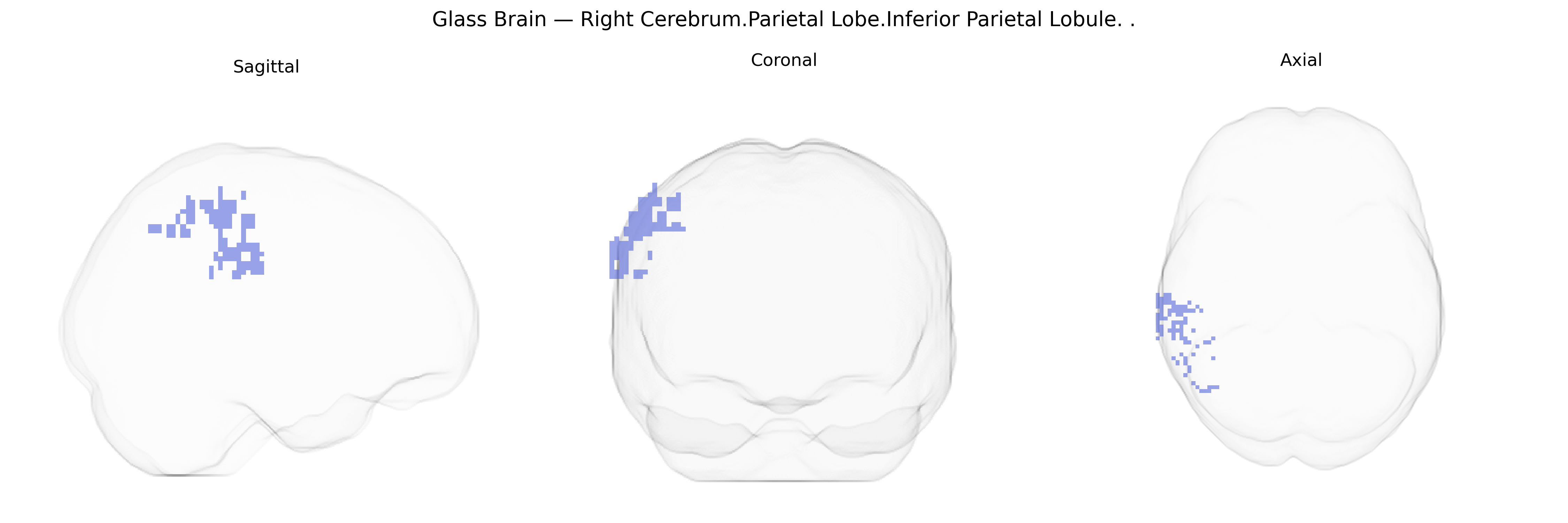
The bilateral Right Cerebrum.Parietal Lobe.Inferior Parietal Lobule corresponds to a portion of the inferior parietal cortex situated in the posterior parietal lobe, wrapping around the posterior end of the Sylvian fissure and bordering the temporal and occipital lobes. This region, encompassing parts of the supramarginal and angular gyri, is implicated in multisensory integration, spatial attention, body-awareness, language processes (including reading and semantic processing), numerical cognition, and aspects of social cognition such as theory of mind. It receives convergent visual, auditory, and somatosensory input and plays a key role in constructing internal representations of space and actions; lesions in this area can lead to deficits such as apraxia, neglect, or language disturbances depending on hemisphere and subregion. There is no direct Wikipedia article for this specific Talairach label; a closely related structure is the Inferior parietal lobule.
Genetic associations involving the bilateral right inferior parietal lobule (IPL) implicate this region in a range of cognitive and neuropsychiatric traits, although most findings come from imaging genetics and large-scale GWAS of brain structure and function rather than region-specific studies keyed exactly to the Talairach 2 mm atlas label. Variants in genes involved in synaptic plasticity and neuronal migration (e.g., BDNF, CNTNAP2, DISC1, and CACNA1C) have been linked to parietal cortical thickness and surface area, as well as to functional activation in the IPL during tasks of attention, working memory, and social cognition. Large GWAS of cortical morphology (e.g., ENIGMA and UK Biobank consortia) have identified multiple loci associated with parietal lobe measures, including regions near genes such as RSPO3, TBR1, and MEF2C, which influence neurodevelopment and synaptic function, and these structural variants correlate with educational attainment, general cognitive performance, and risk for disorders such as schizophrenia, ADHD, and autism spectrum disorder. Imaging genetics studies in Alzheimer’s disease and frontotemporal dementia have reported that APOE ε4 and other AD risk variants are associated with altered IPL volume, cortical thinning, and disrupted default mode network connectivity, reflecting the IPL’s role in episodic memory and spatial–attentional processing. Additional GWAS of functional connectivity and task-evoked activity suggest that polygenic scores for intelligence, reading ability, and mathematical skills modulate activation and connectivity of the IPL within frontoparietal and language networks, while polygenic risk for depression and anxiety shows subtler associations with right IPL structure and resting-state patterns, underscoring this region as a genetically sensitive hub for higher-order cognition and vulnerability to neuropsychiatric disease.
Overview generated by GPT-4o (2026).
Region ID: 889
Hemisphere: bilateral
Atlas: Talairach labels 2mm
Full Quality Version: Download MP4
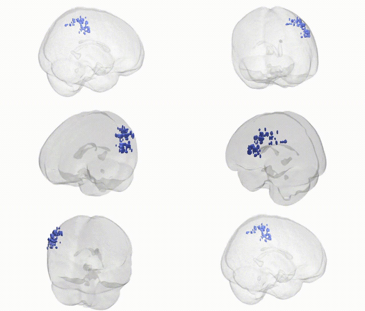
Full Quality Version: Download MP4
Wali Sidiqyar*, Gaurav Rudravaram*, Elyssa M. McMaster, Trent M. Schwartz, Adam M. Saunders, Kurt G. Schilling, Bennett A. Landman "Introducing SPINS: A Shared Public Visualization Library of Neuroanatomical Structures." Medical Imaging with Deep Learning- short paper
This resource is licensed under CC0 1.0 Universal (Public Domain).