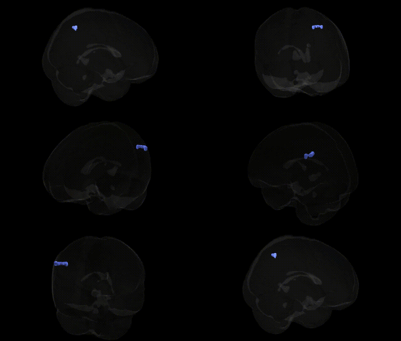
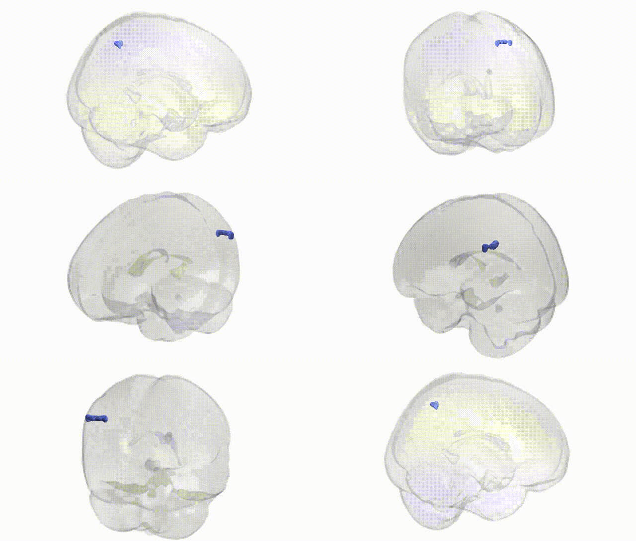
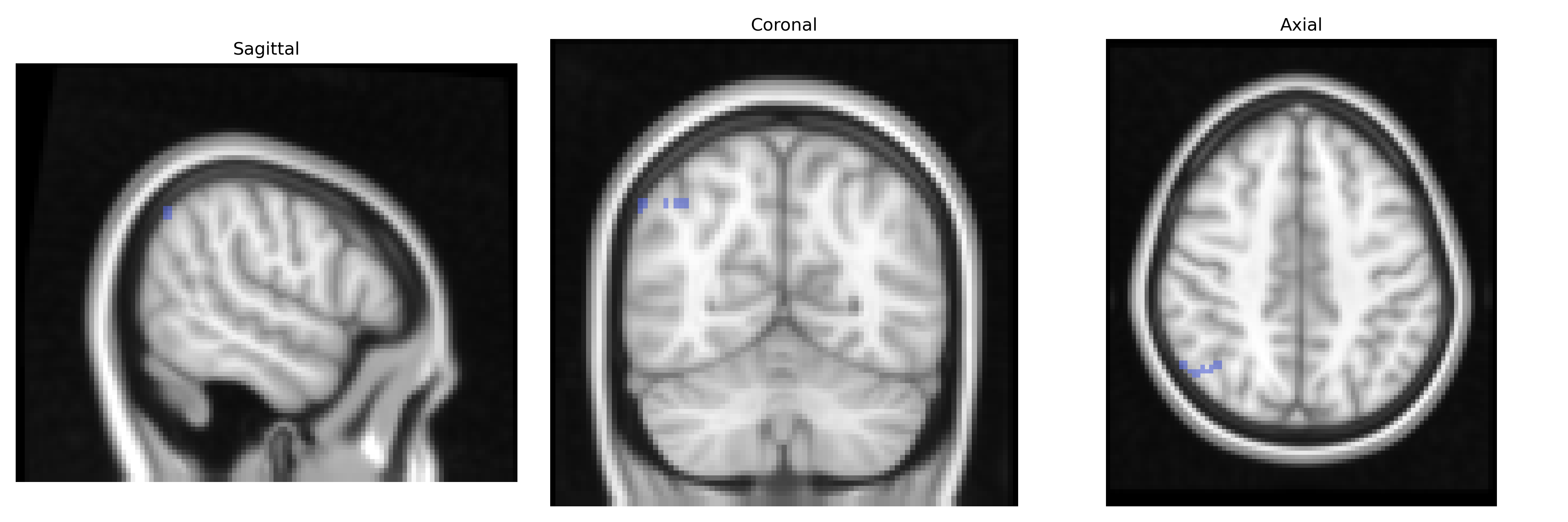
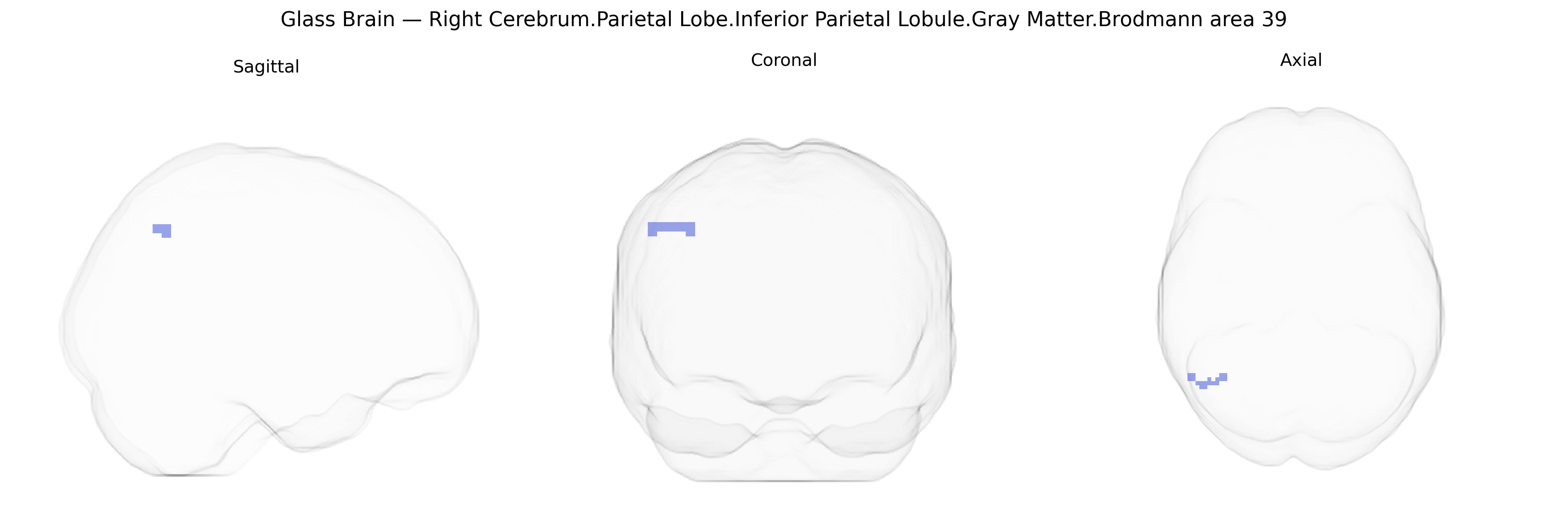
The bilateral Right Cerebrum.Parietal Lobe.Inferior Parietal Lobule.Gray Matter.Brodmann area 39 corresponds to the angular gyrus region within the inferior parietal lobule of the parietal cortex. Brodmann area 39 is a higher-order association area that integrates visual, auditory, and somatosensory information, and is critically involved in language-related functions (such as reading and semantic processing), number comprehension, spatial cognition, and aspects of theory of mind and social cognition. It forms part of the default mode and language networks, with strong connectivity to temporal and frontal association cortices, and shows lateralization of function, particularly in the dominant (usually left) hemisphere, though the Talairach label here refers to the homologous right-sided gray matter region. Angular gyrus
The bilateral right inferior parietal lobule (IPL), Brodmann area 39, has been implicated in multiple genetic and GWAS findings that link its gray matter structure and function to diverse neurocognitive traits and psychiatric disorders. Imaging genetics studies and large-scale consortia such as ENIGMA and UK Biobank have shown that common variants in genes influencing neurodevelopment and synaptic plasticity (e.g., BDNF, CACNA1C, DISC1, MAPT, and several loci in Wnt and axon guidance pathways) are associated with IPL cortical thickness and surface area, as well as parietal lobe gray matter volume more broadly. GWAS of cognitive abilities, educational attainment, and reading/language-related traits have repeatedly implicated parietal association cortices, including BA39, in networks subserving semantic processing, number cognition, attention, and working memory, with significant SNP heritability for IPL morphology and connectivity. Moreover, genetic risk for schizophrenia, bipolar disorder, major depressive disorder, autism spectrum disorder, and ADHD has been associated with structural and functional alterations in the right IPL, including reduced gray matter volume, altered default mode and frontoparietal network connectivity, and atypical lateralization patterns; polygenic risk scores in these conditions often predict parietal lobe measures in neuroimaging cohorts. The right BA39 region also appears in GWAS and imaging genetics studies of Alzheimer’s disease and other dementias, where APOE and other AD-risk loci relate to atrophy and hypometabolism in temporoparietal association cortex, as well as in studies of chronic pain, spatial neglect, and social cognition, underscoring a broad genetic contribution to IPL structure and function across cognitive, neurodevelopmental, and neurodegenerative phenotypes.
Overview generated by GPT-4o (2026).
Region ID: 1015
Hemisphere: bilateral
Atlas: Talairach labels 2mm
Full Quality Version: Download MP4
Full Quality Version: Download MP4
Wali Sidiqyar*, Gaurav Rudravaram*, Elyssa M. McMaster, Trent M. Schwartz, Adam M. Saunders, Kurt G. Schilling, Bennett A. Landman "Introducing SPINS: A Shared Public Visualization Library of Neuroanatomical Structures." Medical Imaging with Deep Learning- short paper
This resource is licensed under CC0 1.0 Universal (Public Domain).