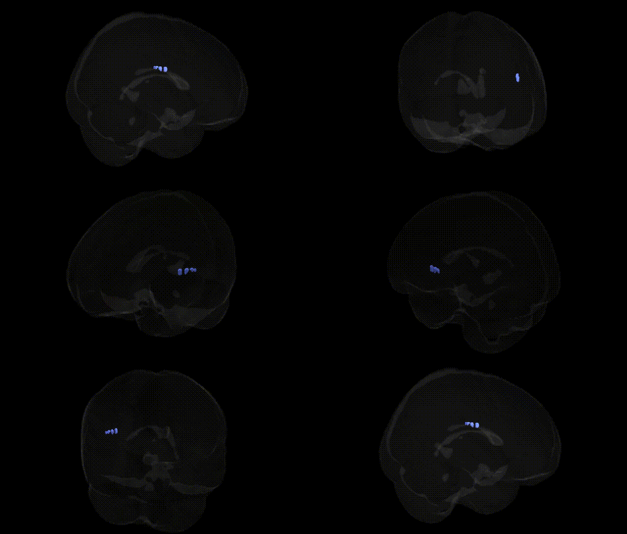
The bilateral Right Cerebrum.Parietal Lobe.Insula.White Matter refers to the subcortical myelinated fiber tracts underlying the insular cortex located deep within the lateral sulcus, at the junction of the parietal, frontal, and temporal lobes on the right hemisphere. These white matter pathways interconnect the insula with widespread cortical and subcortical regions, including parietal association areas, frontal executive regions, limbic structures, and thalamic nuclei, supporting functions such as interoception, multimodal sensory integration, emotional processing, and aspects of autonomic regulation. While the Talairach label emphasizes its parietal lobe adjacency, the insular white matter forms part of a broader insular network involved in salience detection and the coordination of cognitive and affective states. There is no direct Wikipedia article for this precise white matter label; a closely related structure is the Insular cortex.
Genetic associations specifically targeting the bilateral Right Cerebrum.Parietal Lobe.Insula.White Matter region, as defined in the Talairach 2 mm atlas, are not directly reported in the literature, but multiple GWAS and imaging-genetics studies implicate nearby parietal and insular cortices and their underlying white matter in a range of traits and disorders. Common variants near genes involved in axonal growth and myelination (e.g., NTRK1/2, CNTN4, LINGO1, MAG) have been associated with global and regional white matter microstructure (fractional anisotropy, mean diffusivity) in large cohorts such as UK Biobank, which include tracts traversing parietal and insular regions. Parietal and insular white matter integrity has been genetically correlated with cognitive performance (general intelligence, processing speed), educational attainment, and risk for neuropsychiatric conditions including schizophrenia, bipolar disorder, major depressive disorder, and autism spectrum disorder, often via polygenic scores that predict reduced integrity in frontoparietal and insular networks. GWAS of stroke and small-vessel disease (e.g., variants near COL4A1/2, PITX2, ZFHX3) and of white matter hyperintensities implicate vascular and extracellular matrix genes in damage to parietal-insular white matter, while migraine and chronic pain GWAS (involving genes such as TRPM8, LRP1) overlap with structural and functional changes in insular-parietal pathways involved in pain processing and interoception. Although no study isolates this Talairach-defined label as a discrete genetic target, convergent evidence from large-scale imaging-genetics consortia indicates that polygenic influences on connectivity, cognition, mood, and vascular risk are expressed in the white matter underlying parietal and insular cortices, making this region part of distributed networks affected by common and rare genetic variation rather than a uniquely and independently characterized locus.
Overview generated by GPT-4o (2026).
Region ID: 777
Hemisphere: bilateral
Atlas: Talairach labels 2mm
Full Quality Version: Download MP4
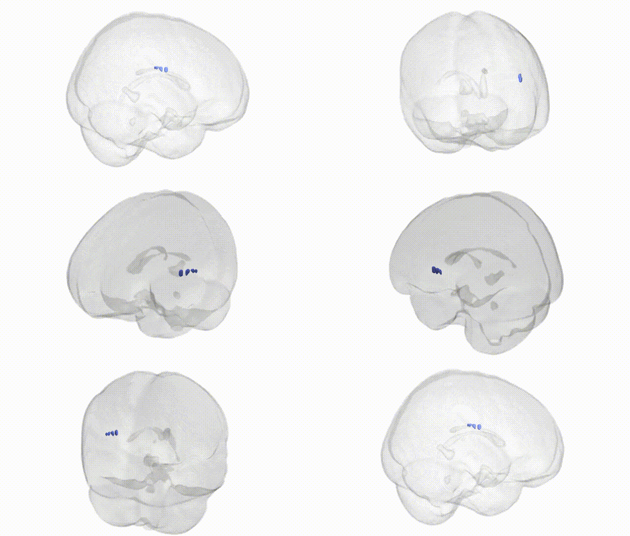
Full Quality Version: Download MP4
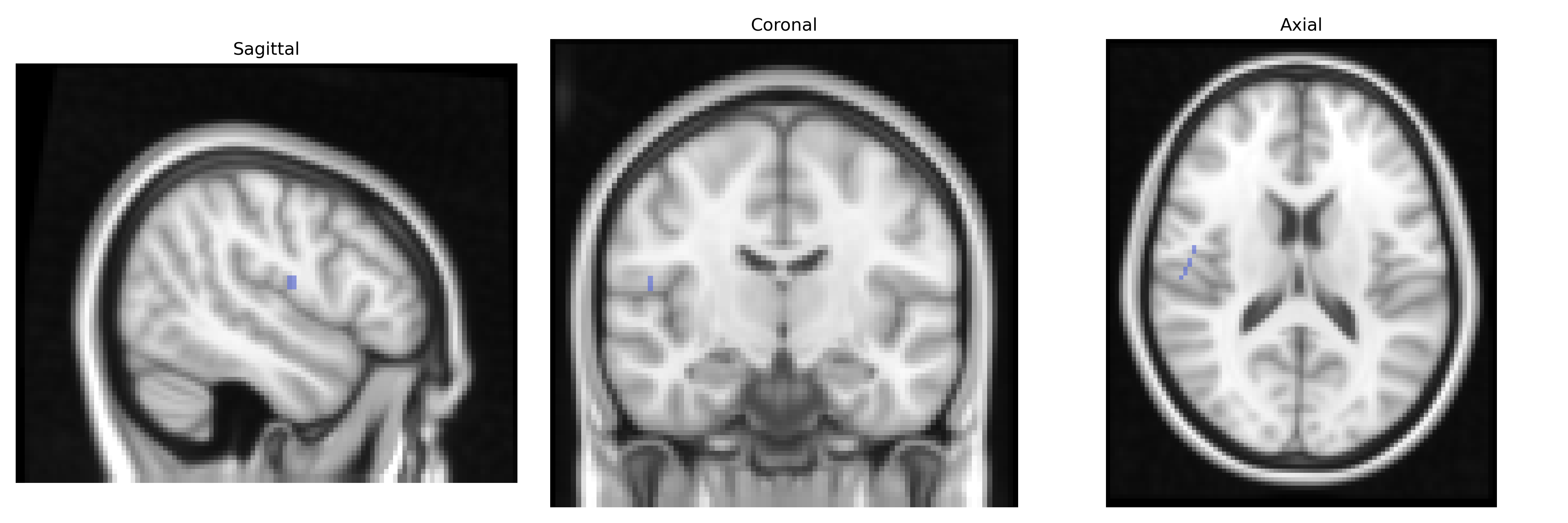
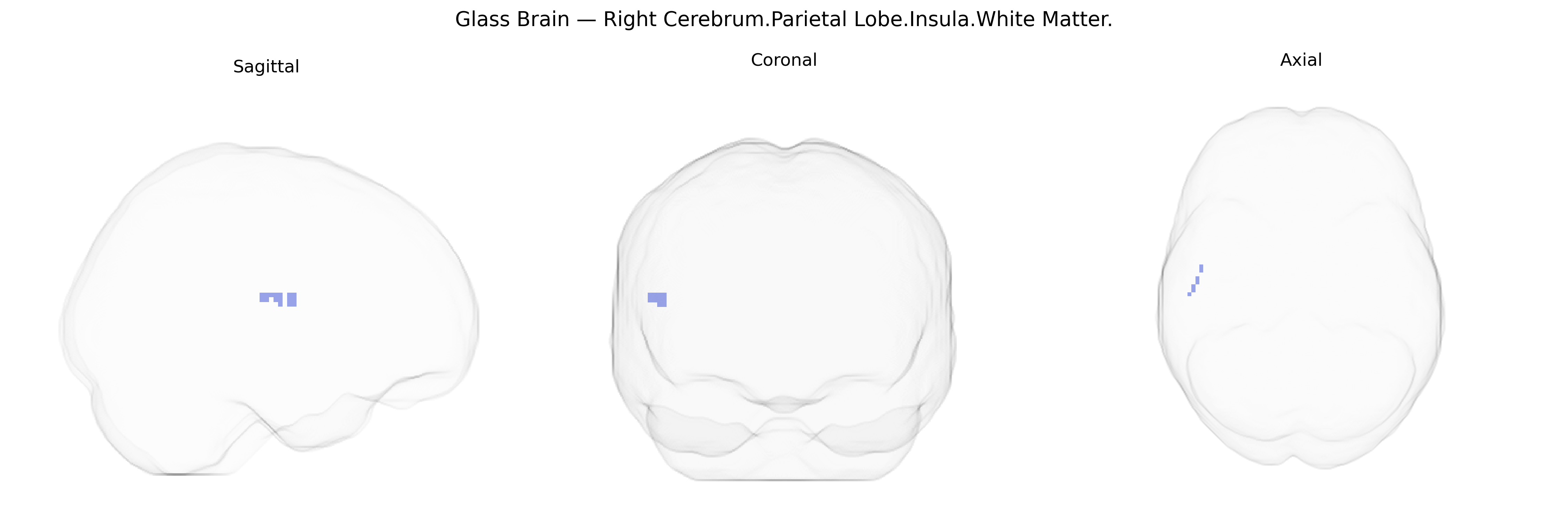
Wali Sidiqyar*, Gaurav Rudravaram*, Elyssa M. McMaster, Trent M. Schwartz, Adam M. Saunders, Kurt G. Schilling, Bennett A. Landman "Introducing SPINS: A Shared Public Visualization Library of Neuroanatomical Structures." Medical Imaging with Deep Learning- short paper
This resource is licensed under CC0 1.0 Universal (Public Domain).