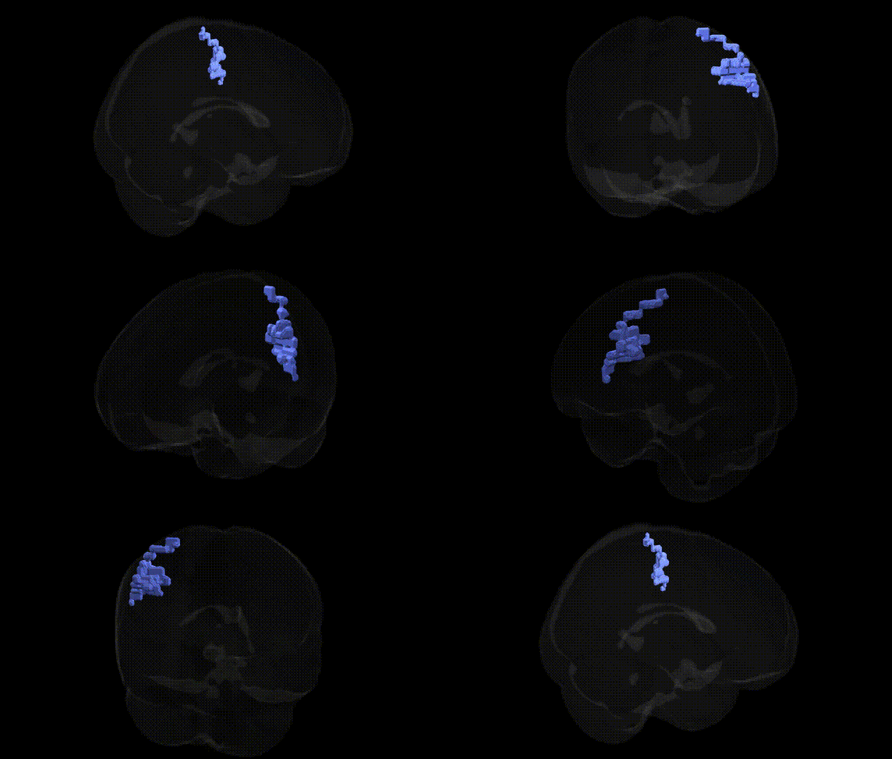
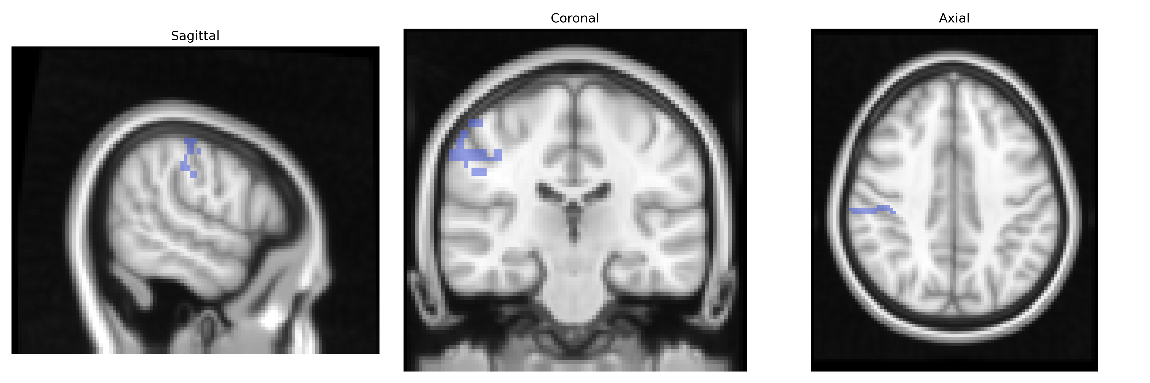
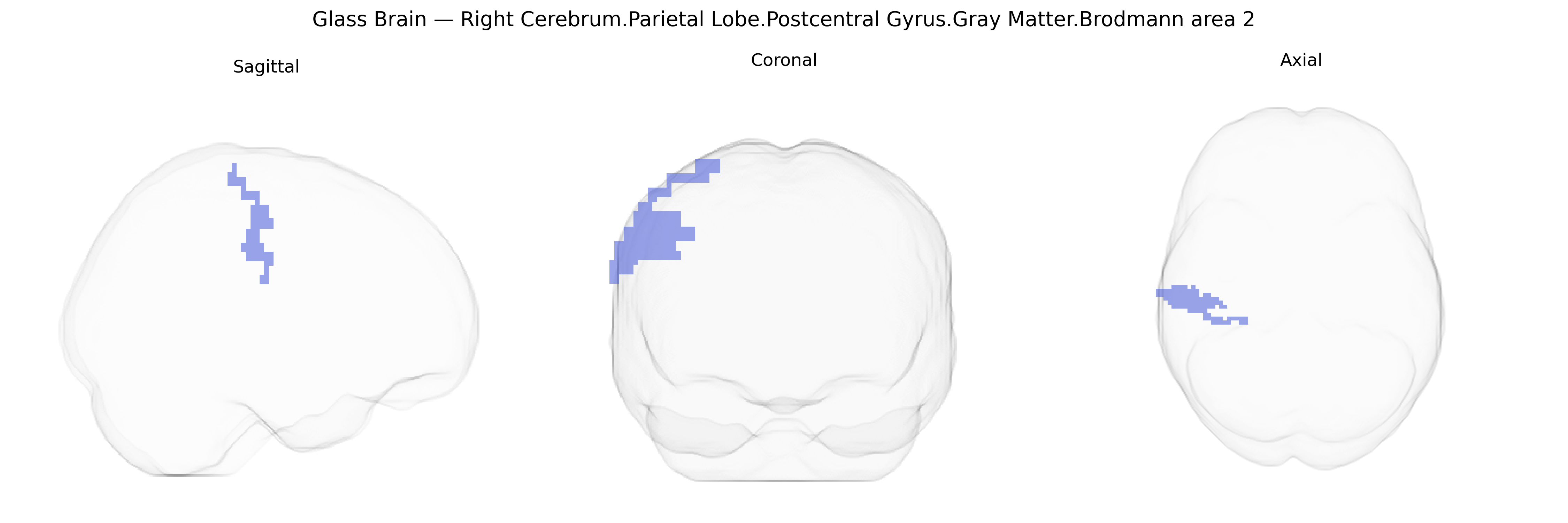
Bilateral Right Cerebrum Parietal Lobe Postcentral Gyrus Gray Matter Brodmann area 2 is a somatosensory cortical region located in the dorsal portion of the postcentral gyrus, adjacent to primary somatosensory cortex (area 3b and area 1). It participates in higher-order processing of tactile and proprioceptive information, integrating inputs from cutaneous and deep receptors to support perception of shape, size, and texture, as well as complex aspects of haptic object recognition. Neurons in Brodmann area 2 exhibit modality convergence and larger, more complex receptive fields than those in more primary somatosensory areas, reflecting its role in constructing integrated sensory representations of the body and external objects. Functionally, this region contributes to sensorimotor coordination and fine manual manipulation by providing detailed feedback about limb position and contact forces. There is no direct Wikipedia article for Brodmann area 2; see the related region Primary somatosensory cortex.
The bilateral right postcentral gyrus gray matter in Brodmann area 2, a primary somatosensory region involved in tactile and proprioceptive processing, has been implicated in several genetic and GWAS-based findings through structural and functional imaging genetics, though often at the level of the broader postcentral gyrus or parietal somatosensory cortex rather than this Talairach-defined parcel specifically. Large brain morphology GWAS consortia (e.g., ENIGMA, UK Biobank) have identified common variants near genes such as HMGA2, IGF1, KIF17, and several neurodevelopmental loci that influence cortical thickness and surface area in the postcentral gyrus, reflecting genetic control of somatosensory cortical development and maturation. Neurodevelopmental and neuropsychiatric disorder studies link variants in synaptic and neurogenesis genes (e.g., CNTNAP2, NRXN1, DISC1-related pathways) to altered postcentral/parietal somatosensory morphology or activation patterns in conditions such as autism spectrum disorder, ADHD, and schizophrenia, where atypical sensory processing and parietal integration are frequent findings. Pain- and touch-related GWAS, including studies of chronic pain, migraine, and pain sensitivity, have reported associations between alleles in nociception and inflammatory pathway genes (e.g., COMT, TRPV1-related networks, cytokine signaling loci) and functional or structural differences in somatosensory cortices that encompass BA2, consistent with a genetic contribution to individual differences in somatosensory representation and pain perception. Motor and sensorimotor trait GWAS, such as those examining hand dexterity, motor ability, or coordination, have also implicated parietal and postcentral regions, with variants in axon guidance, myelination, and synaptic plasticity genes contributing to variability in sensorimotor cortex organization. Overall, genetic studies support a polygenic, distributed architecture in which variants affecting cortical development, synaptic function, and sensory pathways contribute to interindividual differences in structure and function of Brodmann area 2, and to susceptibility for disorders characterized by altered somatosensory and parietal processing, even though direct gene–region mapping at the fine-grained Talairach 2 mm label is not yet routinely reported.
Overview generated by GPT-4o (2026).
Region ID: 892
Hemisphere: bilateral
Atlas: Talairach labels 2mm
Full Quality Version: Download MP4
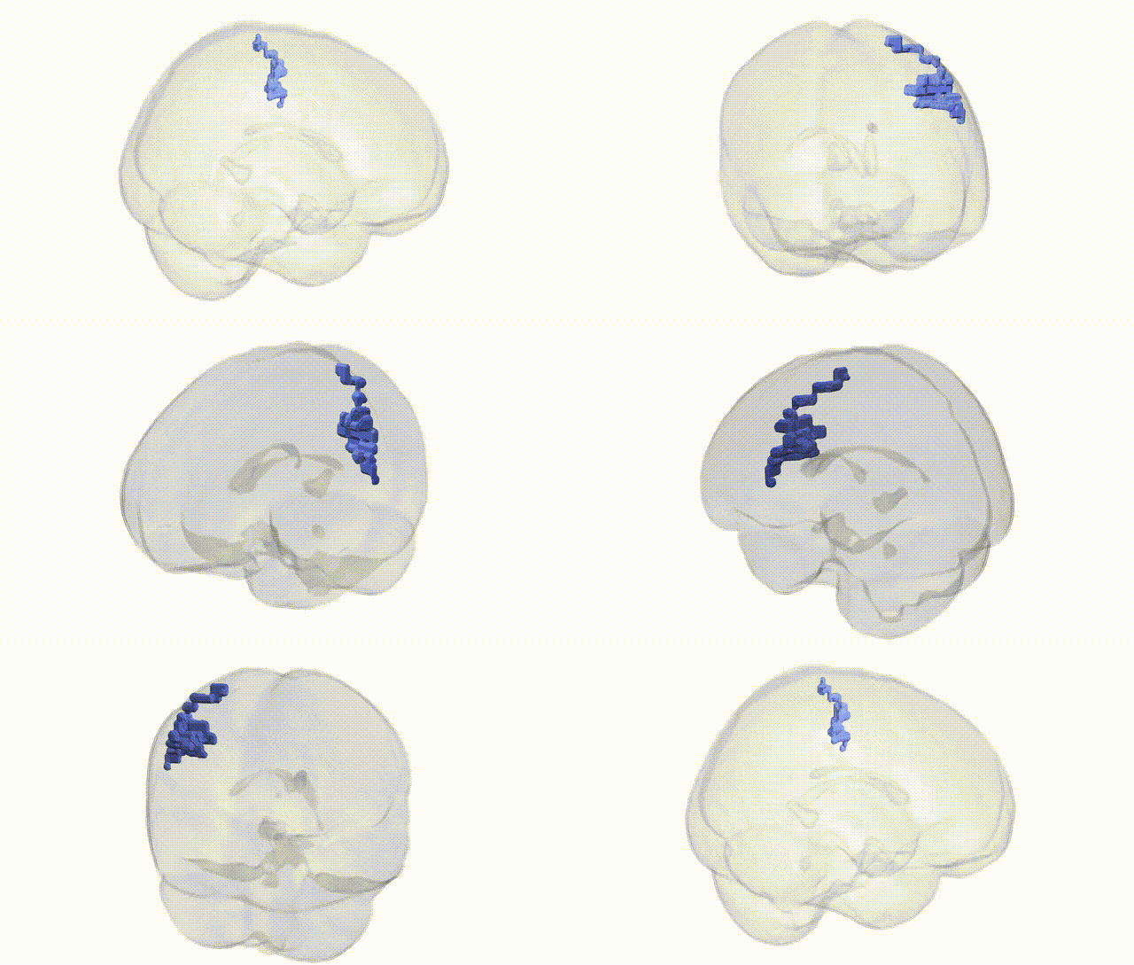
Full Quality Version: Download MP4
Wali Sidiqyar*, Gaurav Rudravaram*, Elyssa M. McMaster, Trent M. Schwartz, Adam M. Saunders, Kurt G. Schilling, Bennett A. Landman "Introducing SPINS: A Shared Public Visualization Library of Neuroanatomical Structures." Medical Imaging with Deep Learning- short paper
This resource is licensed under CC0 1.0 Universal (Public Domain).