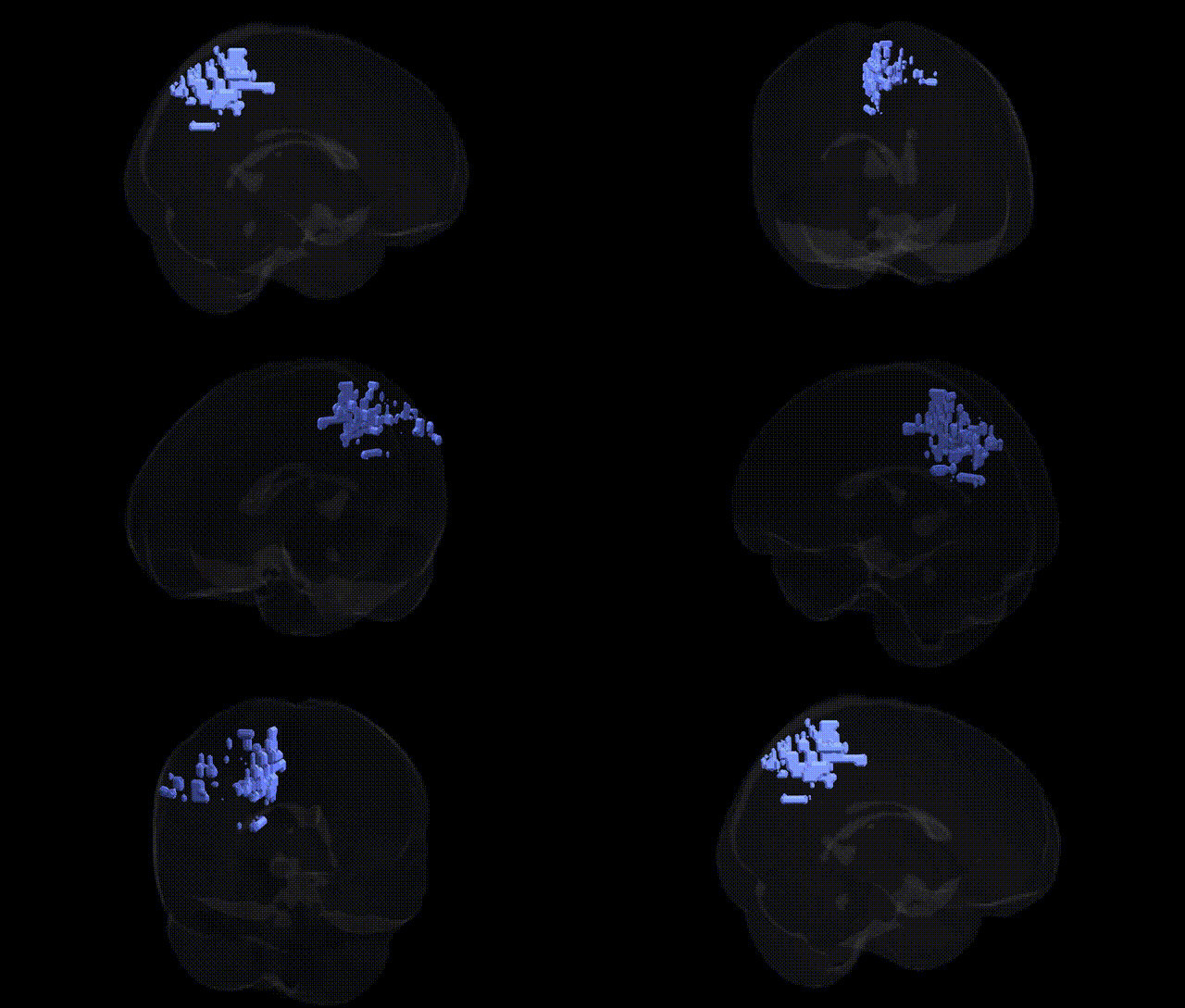
The bilateral Right Cerebrum.Parietal Lobe.Precuneus corresponds to the precuneus region of the superior parietal lobule, situated on the medial surface of the parietal lobe between the marginal branch of the cingulate sulcus and the parieto‑occipital sulcus. It is a heteromodal association area with widespread connectivity to frontal, parietal, temporal, and subcortical structures, and plays key roles in visuo-spatial imagery, self-referential processing, episodic memory retrieval, and aspects of consciousness and default-mode network activity. Functionally, the precuneus participates in integrating sensory information for higher-order representation of space and the body, supports mental imagery and perspective taking, and contributes to internally directed cognitive processes such as mind-wandering and autobiographical memory. Precuneus
The bilateral right precuneus (Right Cerebrum.Parietal Lobe.Precuneus in the Talairach 2 mm atlas) has been implicated in numerous genetic and GWAS findings through its role in default mode network activity, self-referential processing, and visuospatial integration, rather than through variants uniquely tied to this precise parcel. Imaging genetics studies repeatedly link common variants in APOE (especially ε4) and other Alzheimer’s disease–related genes (CLU, PICALM, CR1) to altered precuneus structure, hypometabolism, and amyloid burden, reflecting its early involvement in Alzheimer’s pathology. Large-scale GWAS of cortical thickness and surface area (e.g., ENIGMA and UK Biobank) identify polygenic influences from neurodevelopmental and synaptic genes on parietal/precuneus morphology, with loci such as those near KIAA0586, MIR2113, and others associated with global and parietal cortical measures that encompass this region. Precuneus activity and connectivity show heritable variation and have been associated with schizophrenia and major depression risk via polygenic risk scores and variants in genes involved in glutamatergic signaling and synaptic plasticity, while autism spectrum disorder and ADHD risk variants are linked to altered default mode network connectivity that prominently involves the precuneus. GWAS for cognitive traits including intelligence, memory, and educational attainment implicate gene sets expressed in association cortex (including precuneus) rather than single region-specific genes, and psychiatric and personality GWAS (e.g., neuroticism, risk-taking) converge on distributed networks where the precuneus acts as a hub, indicating that genetic influences on this region largely reflect polygenic effects on broad cortical and network architecture rather than a distinct, precuneus-specific genetic signature.
Overview generated by GPT-4o (2026).
Region ID: 862
Hemisphere: bilateral
Atlas: Talairach labels 2mm
Full Quality Version: Download MP4
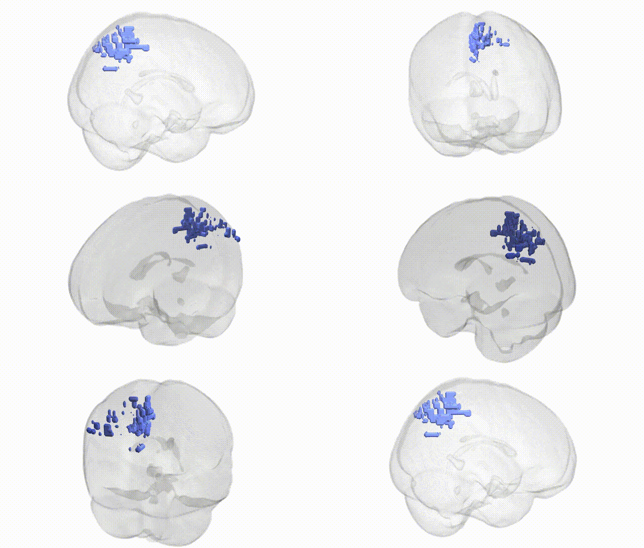
Full Quality Version: Download MP4
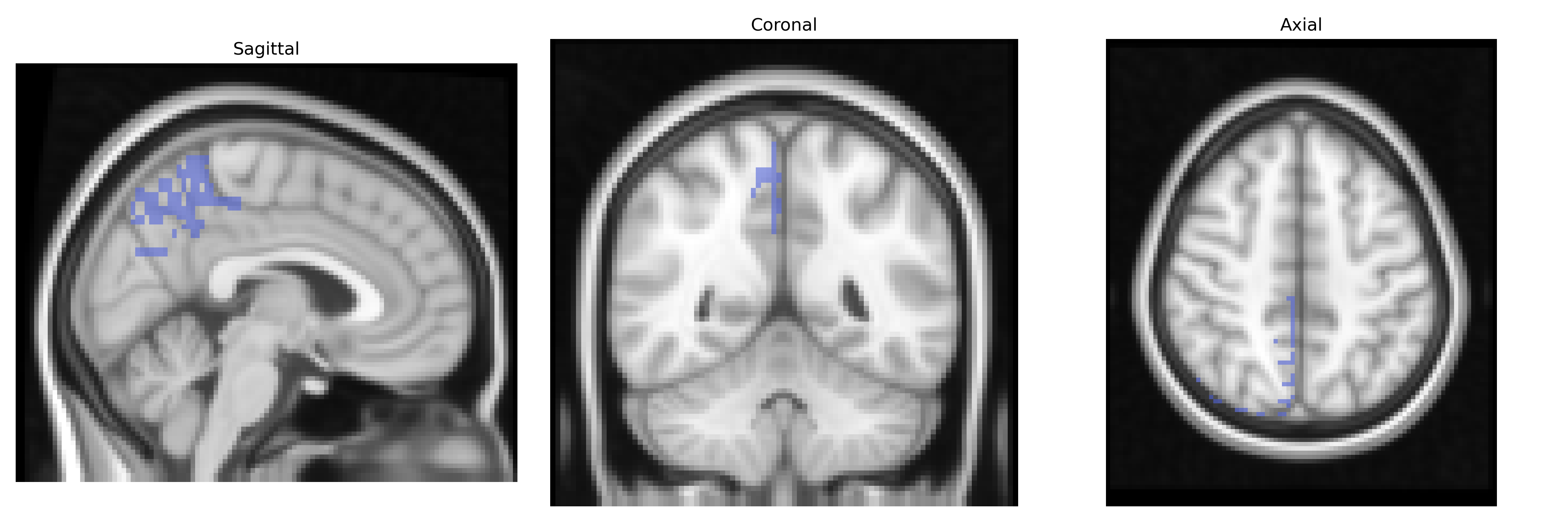
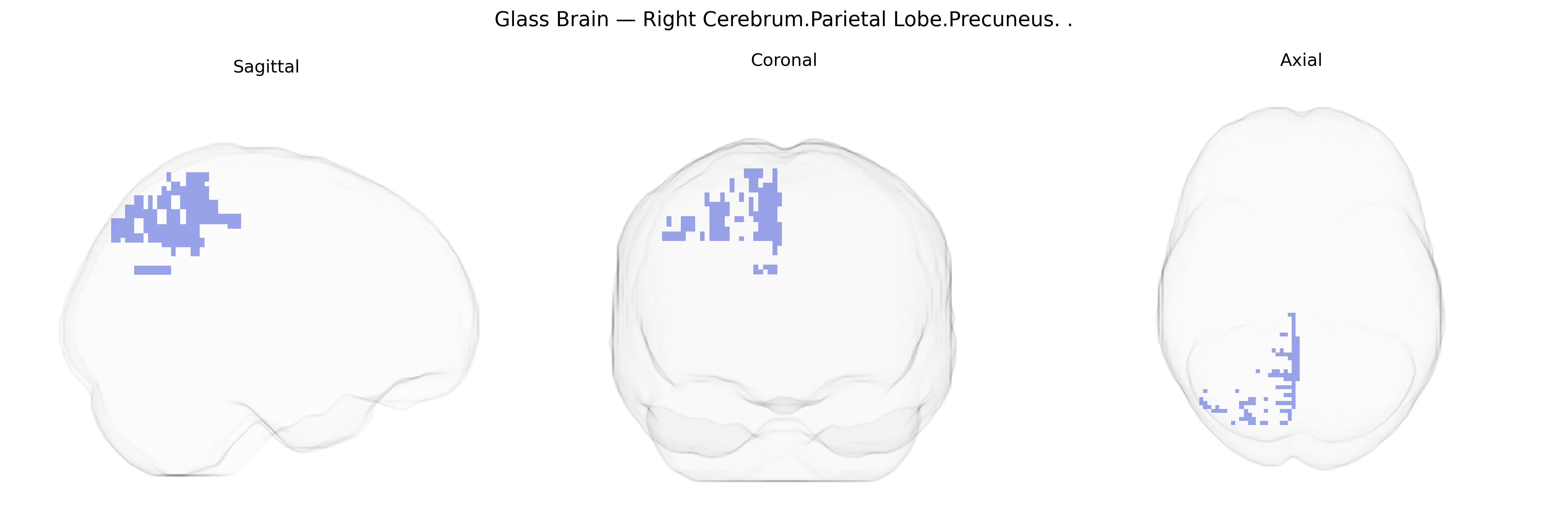
Wali Sidiqyar*, Gaurav Rudravaram*, Elyssa M. McMaster, Trent M. Schwartz, Adam M. Saunders, Kurt G. Schilling, Bennett A. Landman "Introducing SPINS: A Shared Public Visualization Library of Neuroanatomical Structures." Medical Imaging with Deep Learning- short paper
This resource is licensed under CC0 1.0 Universal (Public Domain).