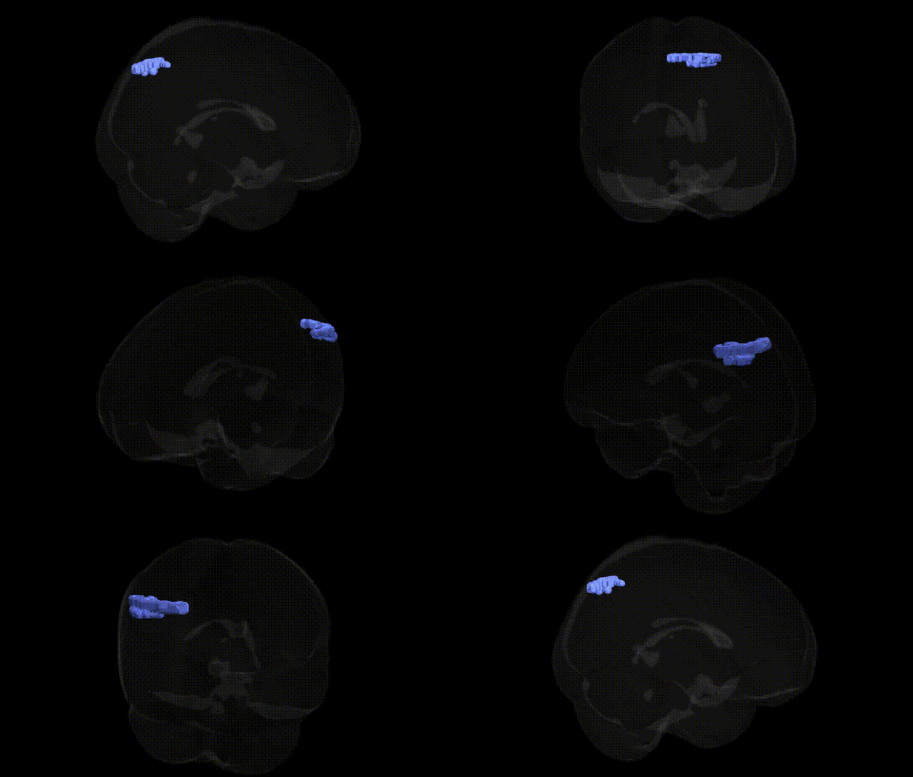
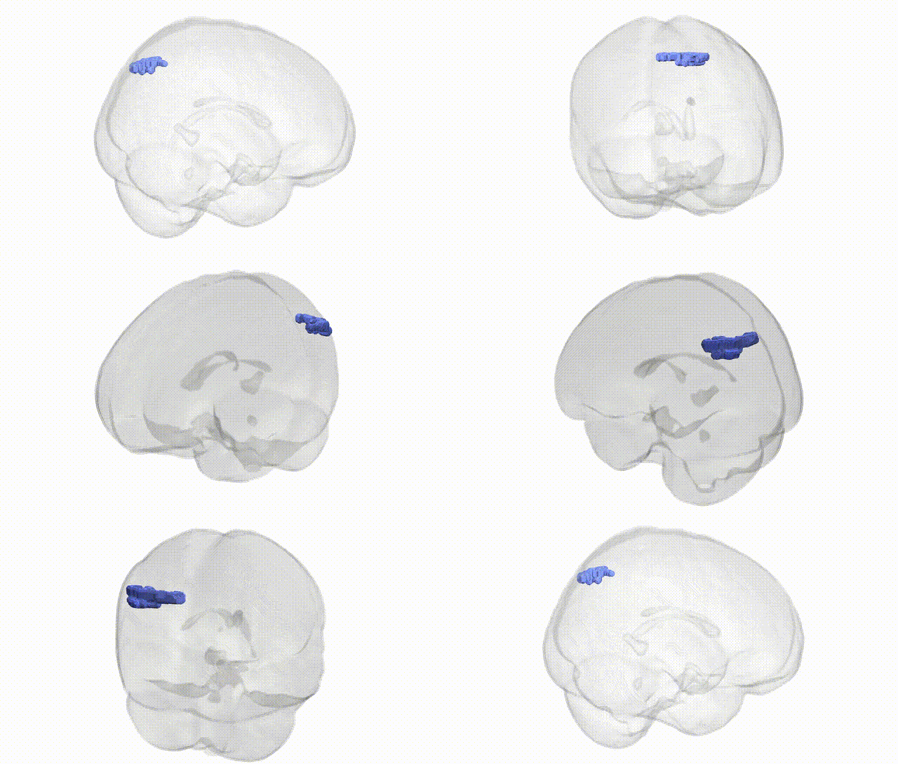
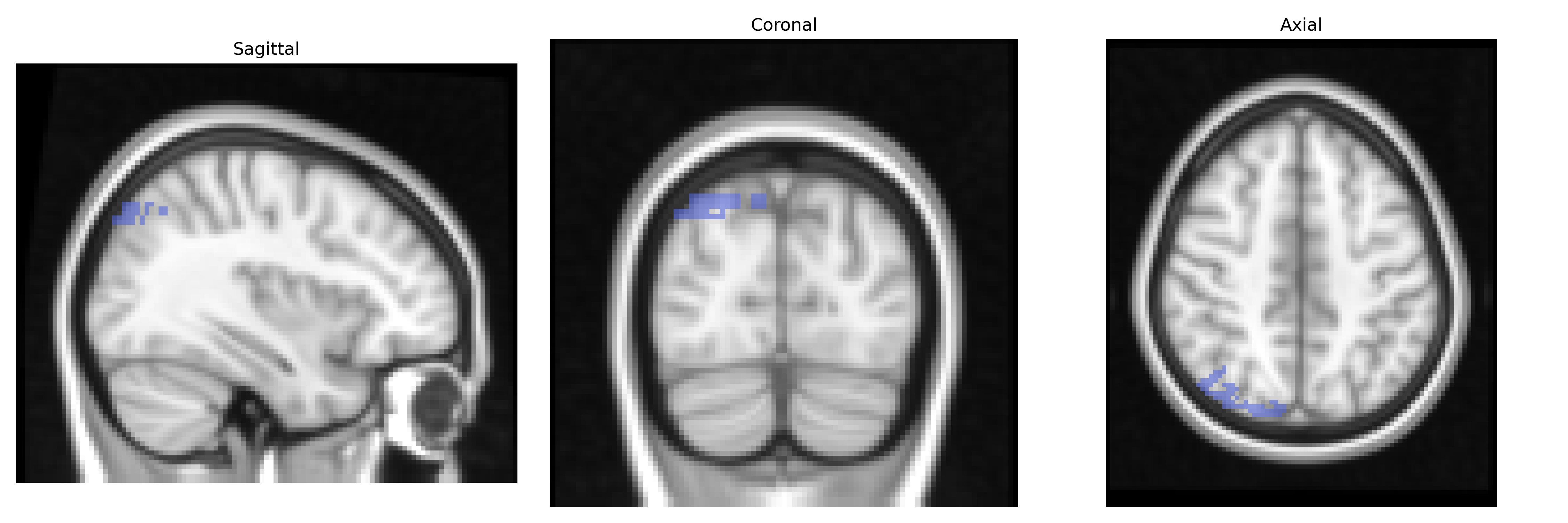
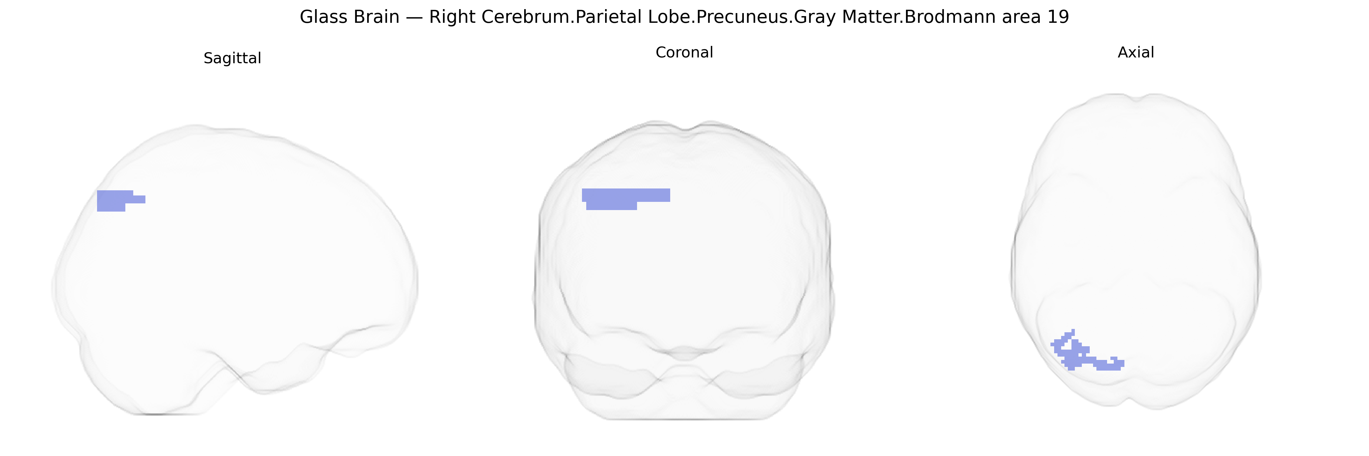
The bilateral Right Cerebrum.Parietal Lobe.Precuneus.Gray Matter.Brodmann area 19 corresponds to an association visual region located medially in the parietal lobe, bordering the occipital cortex and integrated with the precuneus. Brodmann area 19, part of the extrastriate visual cortex, is involved in higher-order visual processing, including integration of visual features, complex pattern recognition, and aspects of visuospatial analysis. Within the precuneus, this territory contributes to multimodal integration of visual information with spatial and self-referential processing, participating in networks related to visual imagery, navigation, and the coordination of perception with internally generated representations. There is no direct Wikipedia article for this exact Talairach-defined region; a closely related structure is Brodmann area 19.
The bilateral right precuneus gray matter in Brodmann area 19, a posterior medial parietal/occipital association region, has been implicated in multiple imaging genetics and GWAS studies through its cortical thickness, surface area, and functional connectivity profiles rather than via region-specific “BA19-precuneus” loci. Large-scale neuroimaging GWAS (e.g., ENIGMA, UK Biobank) consistently highlight polygenic influences on parietal and occipital cortical morphology, with common variants near genes such as HMGA2, MIR9-2, IGF1, and others contributing to global and regional cortical thickness and surface area that include the precuneus/BA19 territory. The precuneus has emerged as a key node in the default mode network (DMN), and genetic variation affecting DMN connectivity—particularly in APOE (ε4), CLU, PICALM, and CR1—has been associated with Alzheimer’s disease risk and with atrophy or hypometabolism in medial parietal regions including the precuneus. Schizophrenia, major depression, and autism GWAS implicate synaptic and neurodevelopmental genes (e.g., CACNA1C, GRIN2A, CNTNAP2) whose expression patterns and imaging correlates often include altered structure or connectivity in the precuneus and adjacent BA19 cortex, especially in tasks involving visuospatial integration, self-referential processing, or social cognition. Polygenic scores for cognitive ability and educational attainment have also been associated with variation in parietal/occipital cortical measures, including the precuneus, suggesting that many small-effect variants collectively influence the structure and function of this region; however, no single gene or variant is uniquely or specifically tied to “bilateral Right Cerebrum.Parietal Lobe.Precuneus.Gray Matter.Brodmann area 19” in the Talairach 2 mm atlas, and current evidence supports a broad, highly polygenic architecture affecting this region as part of larger cortical and network-level phenotypes.
Overview generated by GPT-4o (2026).
Region ID: 987
Hemisphere: bilateral
Atlas: Talairach labels 2mm
Full Quality Version: Download MP4
Full Quality Version: Download MP4
Wali Sidiqyar*, Gaurav Rudravaram*, Elyssa M. McMaster, Trent M. Schwartz, Adam M. Saunders, Kurt G. Schilling, Bennett A. Landman "Introducing SPINS: A Shared Public Visualization Library of Neuroanatomical Structures." Medical Imaging with Deep Learning- short paper
This resource is licensed under CC0 1.0 Universal (Public Domain).