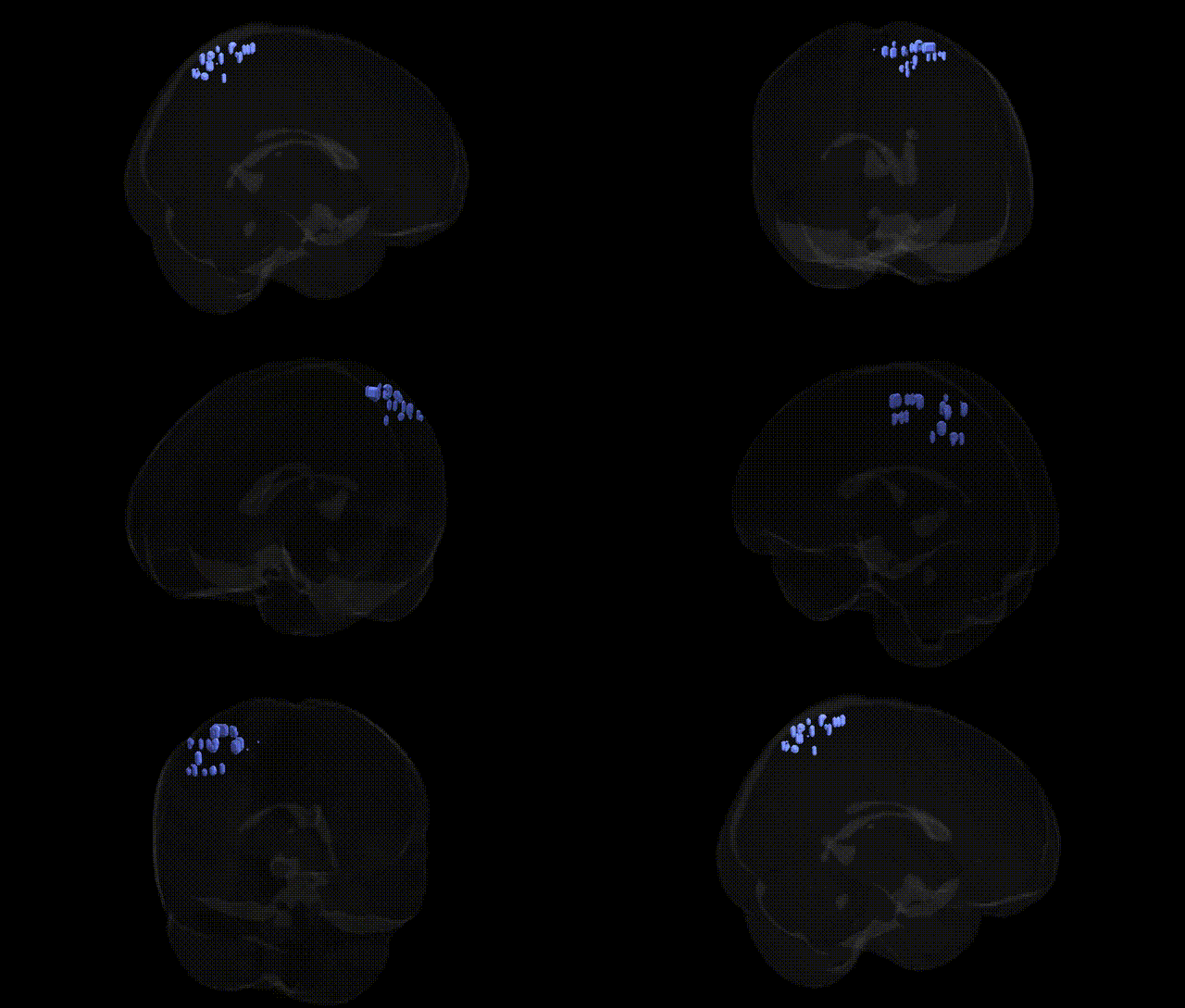
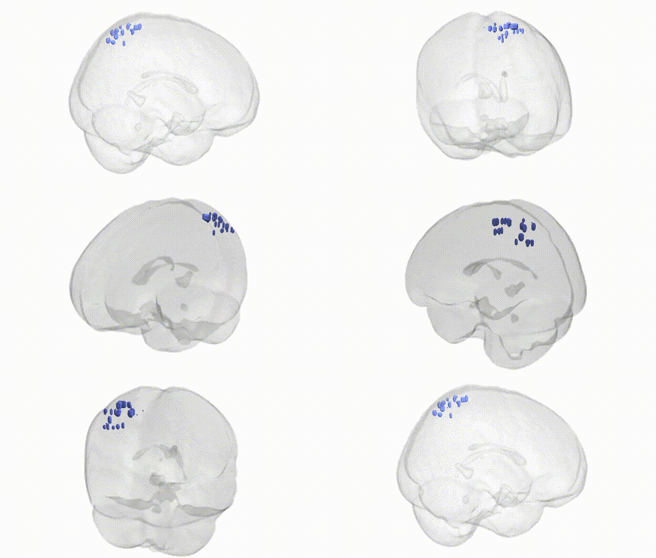
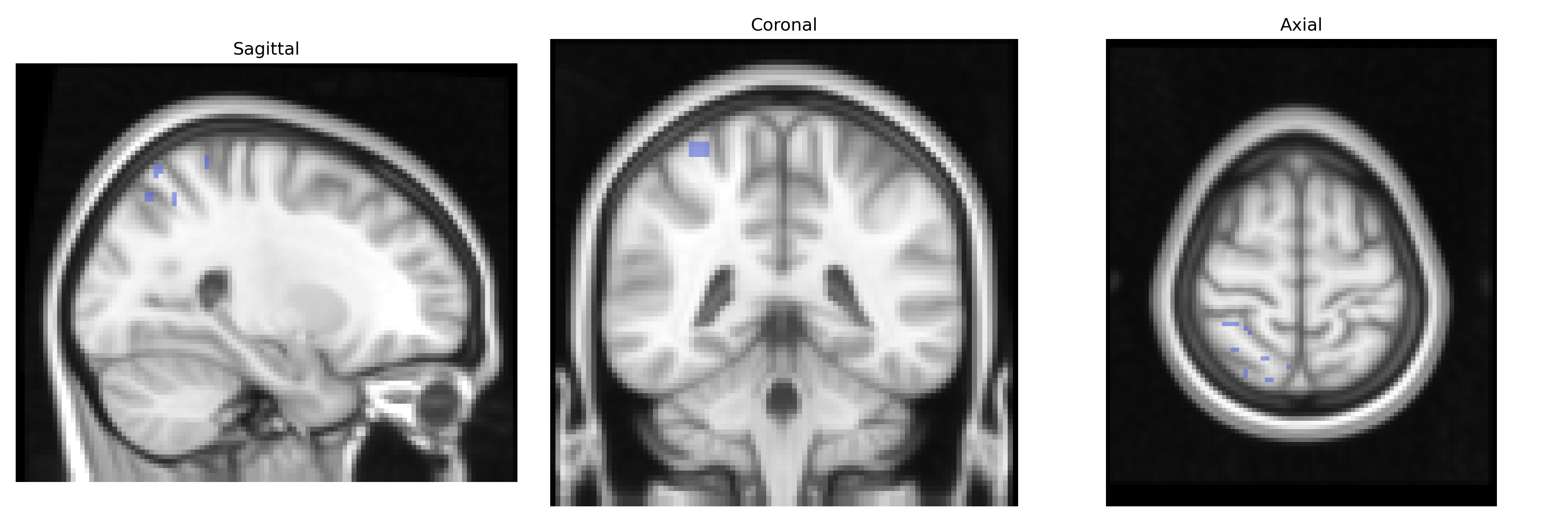
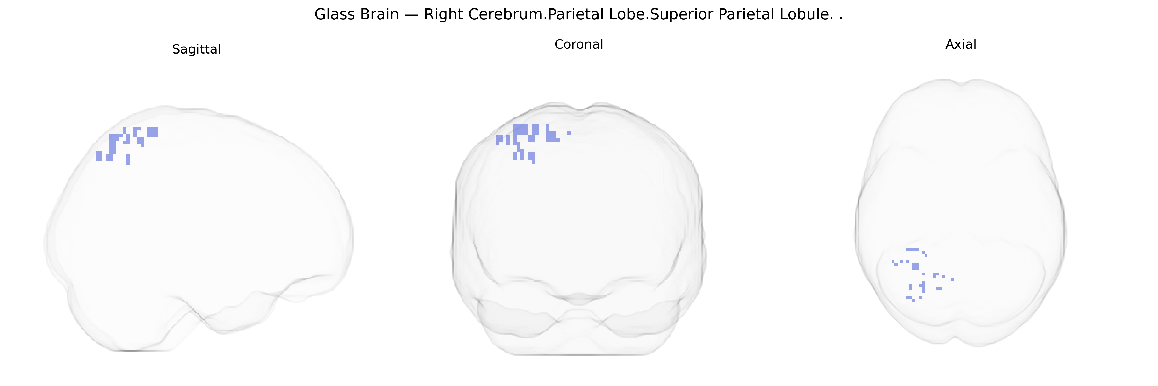
The bilateral Right Cerebrum.Parietal Lobe.Superior Parietal Lobule corresponds to associative cortex located on the dorsal aspect of the parietal lobe, bordering the intraparietal sulcus and extending toward the medial parietal surface. This region is strongly implicated in higher-order somatosensory integration, visuospatial processing, attention, and sensorimotor transformation, contributing to functions such as spatial orientation, body schema representation, and the coordination of visually guided movements. Neuronal populations within the superior parietal lobule integrate inputs from visual, somatosensory, and proprioceptive systems, and are involved in the maintenance and manipulation of information in spatial working memory as well as in the control of reaching and grasping. Lesions in this area can lead to deficits in spatial perception, tactile discrimination, and complex motor coordination, including aspects of apraxia and hemispatial neglect.
The bilateral Right Cerebrum.Parietal Lobe.Superior Parietal Lobule, as defined in the Talairach 2 mm atlas, has been implicated in genetic studies primarily through its roles in visuospatial processing, attention, motor control, and higher-order cognition. GWAS and imaging-genetics work show that cortical thickness, surface area, and functional connectivity of superior parietal regions are influenced by polygenic variation, including loci near genes such as HMGA2, KIAA0586, and others identified in large ENIGMA and UK Biobank analyses of brain morphology. Variants affecting synaptic plasticity, axon guidance, and neurodevelopmental pathways (e.g., in genes like BDNF, CNTNAP2, and FOXP1) have been associated with parietal structure or activation patterns, and these in turn correlate with cognitive performance measures such as fluid intelligence, working memory, and mathematical ability in GWAS of cognition and educational attainment. Disorders with genetic risk that show robust structural or functional alterations in superior parietal cortex include schizophrenia (with risk loci such as ZNF804A and CACNA1C contributing to frontoparietal network changes), ADHD (polygenic burden influencing attention networks and parietal activation), autism spectrum disorder (rare and common variants in synaptic and neurodevelopmental genes linked to atypical parietal connectivity), and Alzheimer’s disease (APOE and other GWAS loci related to parietal atrophy and hypometabolism in later stages). Additionally, genetic influences on visuospatial neglect, motor imagery, and sensorimotor integration have been connected to superior parietal lobule involvement, although these typically reflect distributed network genetics rather than region-specific single-gene effects.
Overview generated by GPT-4o (2026).
Region ID: 1038
Hemisphere: bilateral
Atlas: Talairach labels 2mm
Full Quality Version: Download MP4
Full Quality Version: Download MP4
Wali Sidiqyar*, Gaurav Rudravaram*, Elyssa M. McMaster, Trent M. Schwartz, Adam M. Saunders, Kurt G. Schilling, Bennett A. Landman "Introducing SPINS: A Shared Public Visualization Library of Neuroanatomical Structures." Medical Imaging with Deep Learning- short paper
This resource is licensed under CC0 1.0 Universal (Public Domain).