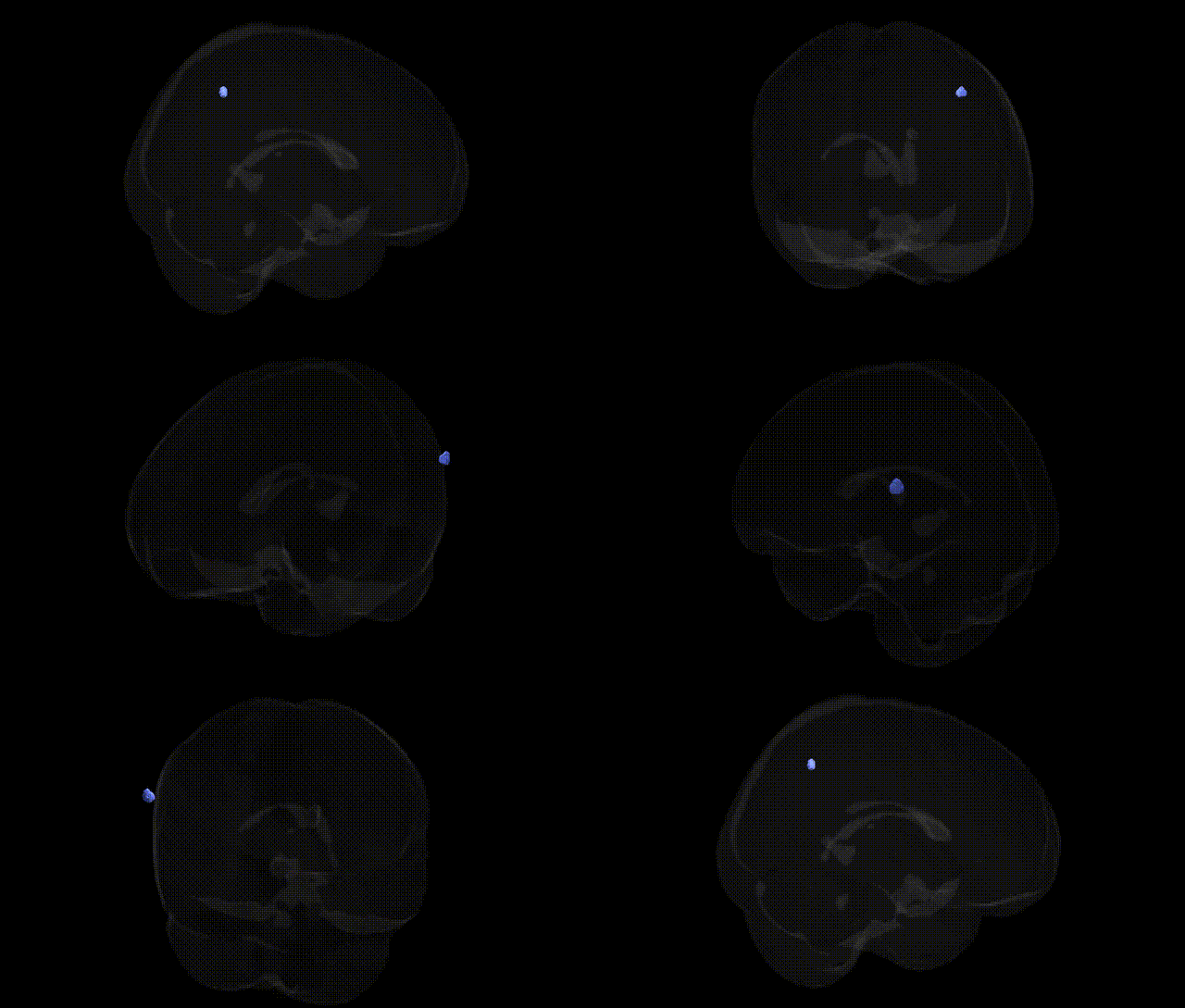
Bilateral Right Cerebrum.Parietal Lobe.Supramarginal Gyrus.Gray Matter.Brodmann area 39 corresponds to the right-hemisphere portions of the angular/supramarginal region within the inferior parietal lobule, as defined in the Talairach 2 mm Atlas. Brodmann area 39 is a multimodal association cortex that integrates visual, auditory, and somatosensory information, contributing critically to language-related processes (such as semantic processing and reading), spatial cognition, number processing, and higher-order conceptual integration. Functionally, this region participates in networks for theory of mind, episodic memory retrieval, and the integration of contextual information, and its disruption is commonly associated with deficits such as alexia, agraphia, and aspects of Gerstmann syndrome. There is no direct Wikipedia article for this exact atlas label; a closely related and encompassing structure is the Angular gyrus.
The right supramarginal gyrus (SMG; Brodmann area 39 in the parietal lobe) has been implicated in multiple genetically influenced traits and disorders, although most genetic findings come from imaging genetics and GWAS of regional cortical measures rather than Talairach-based labels specifically. Large-scale neuroimaging GWAS (e.g., ENIGMA and UK Biobank) have identified heritable variation in cortical thickness and surface area of parietal association regions, including supramarginal/inferior parietal areas, with common variants in genes involved in neuronal development, synaptic signaling, and cytoskeletal architecture (such as variants near genes like CENPO, HMGA2, and others) contributing to inter-individual differences. Functionally, the right SMG participates in language processing (phonological and semantic integration), social cognition/empathy, attention reorientation, and multisensory integration; genetic risk variants for dyslexia and language-related disorders (e.g., in DCDC2, KIAA0319, CNTNAP2) have shown imaging associations with structural and functional changes in temporo-parietal regions that encompass or neighbor BA39. GWAS of autism spectrum disorder, schizophrenia, and attention-deficit/hyperactivity disorder have repeatedly implicated networks including the right inferior parietal/supramarginal region, with polygenic risk scores for these disorders associated with altered gray matter volume or connectivity in BA39 territory, though no single locus is uniquely specific to this area. In social and emotional domains, variants influencing oxytocin and serotonin signaling (e.g., OXTR, SLC6A4) have been linked to differences in empathy and social cognition tasks that recruit the right SMG, and imaging genetics work has associated these alleles with modulation of activation in SMG during perspective-taking and emotional prosody processing. Overall, current evidence indicates that BA39/supramarginal gray matter is a heritable association cortex region whose structure and function reflect polygenic influences from widespread neurodevelopmental and synaptic genes, and it emerges as a consistent node in genetically modulated language, social cognition, and neuropsychiatric risk networks, rather than a region tied to a small set of uniquely specific loci.
Overview generated by GPT-4o (2026).
Region ID: 975
Hemisphere: bilateral
Atlas: Talairach labels 2mm
Full Quality Version: Download MP4
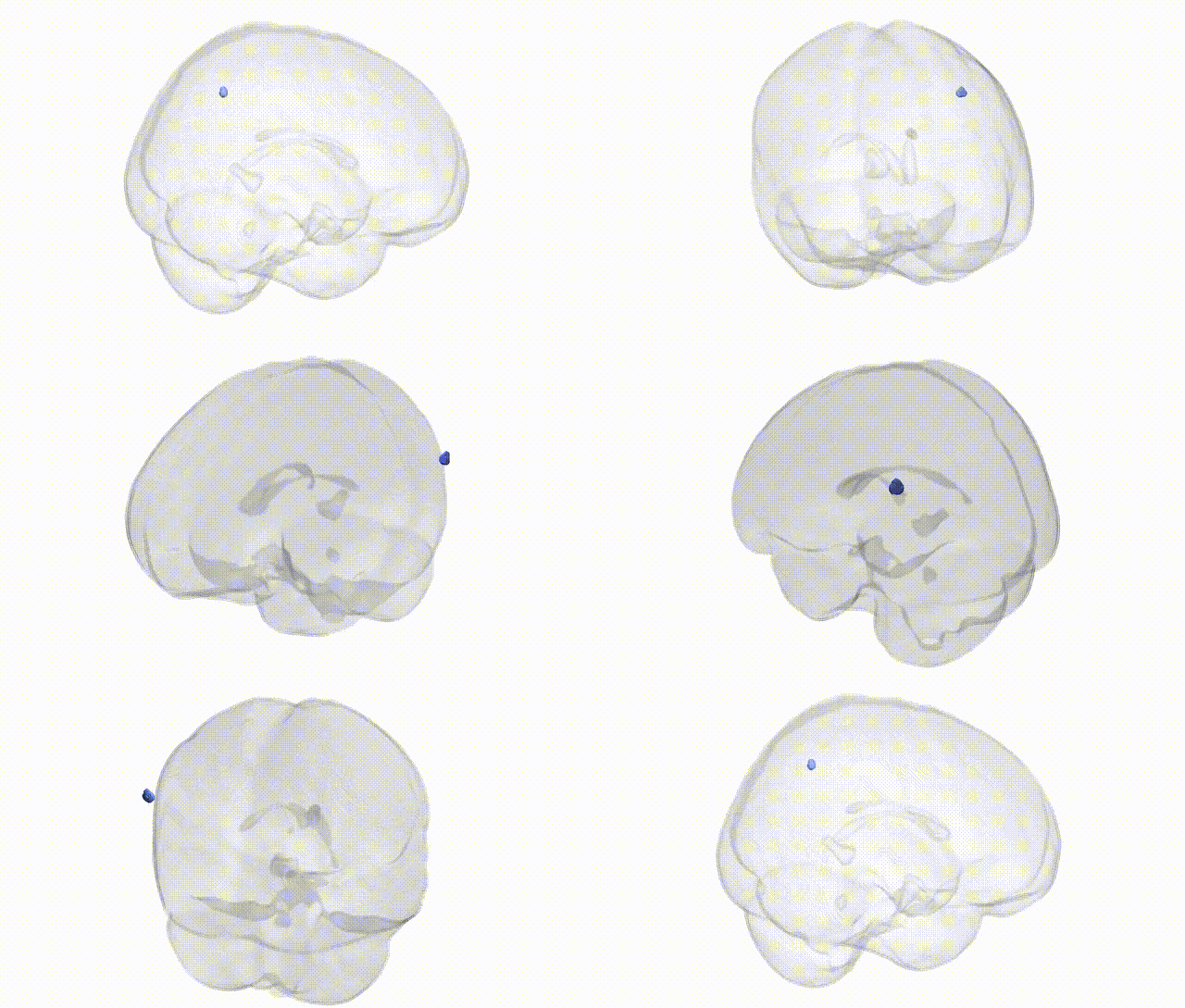
Full Quality Version: Download MP4
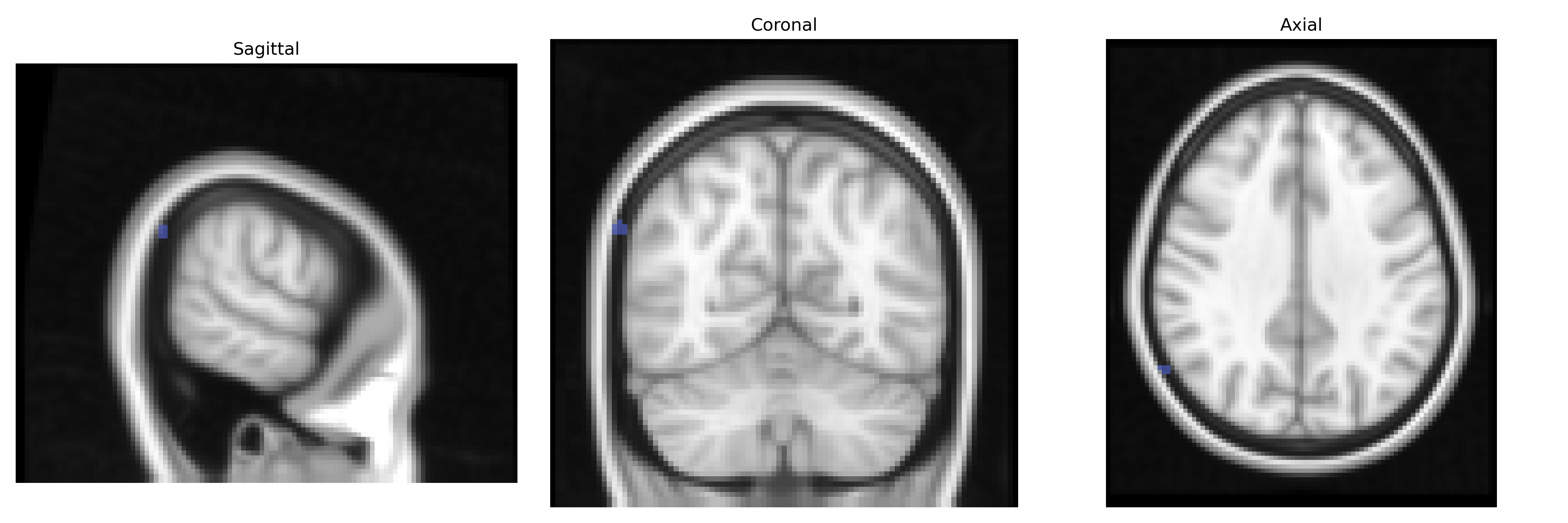
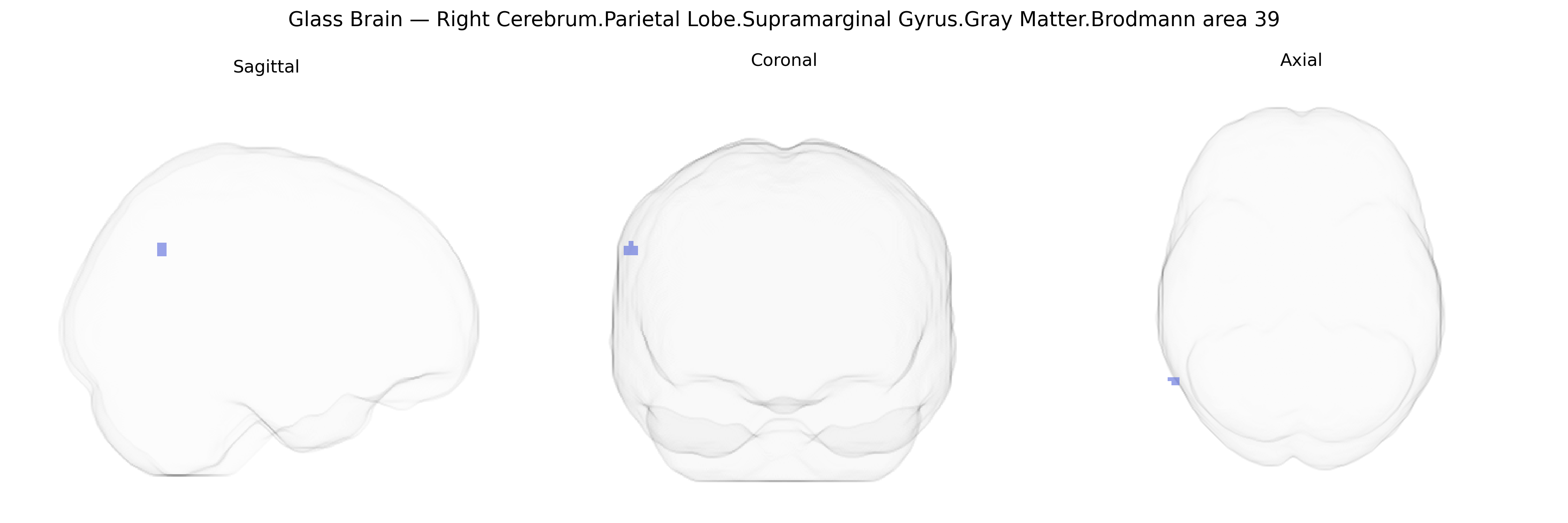
Wali Sidiqyar*, Gaurav Rudravaram*, Elyssa M. McMaster, Trent M. Schwartz, Adam M. Saunders, Kurt G. Schilling, Bennett A. Landman "Introducing SPINS: A Shared Public Visualization Library of Neuroanatomical Structures." Medical Imaging with Deep Learning- short paper
This resource is licensed under CC0 1.0 Universal (Public Domain).