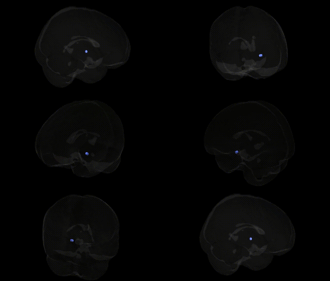
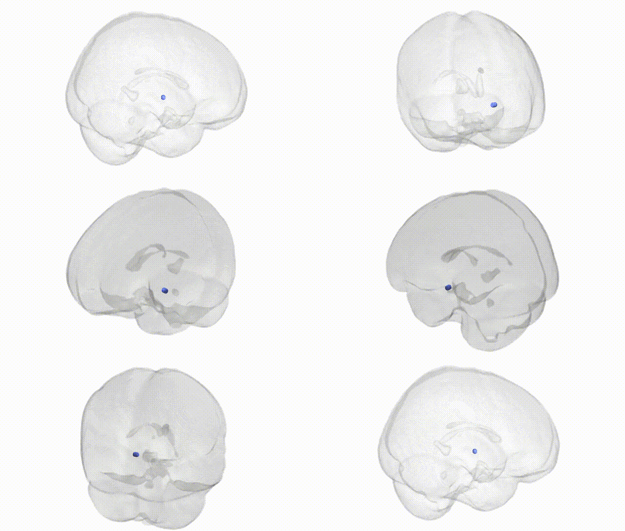
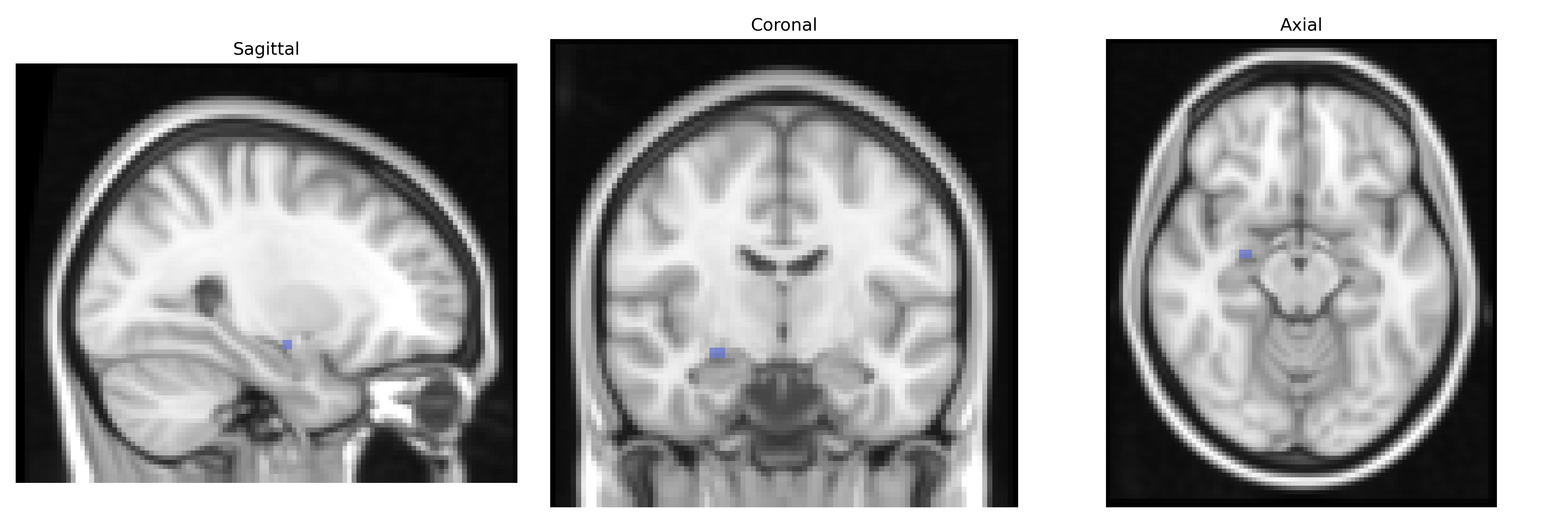
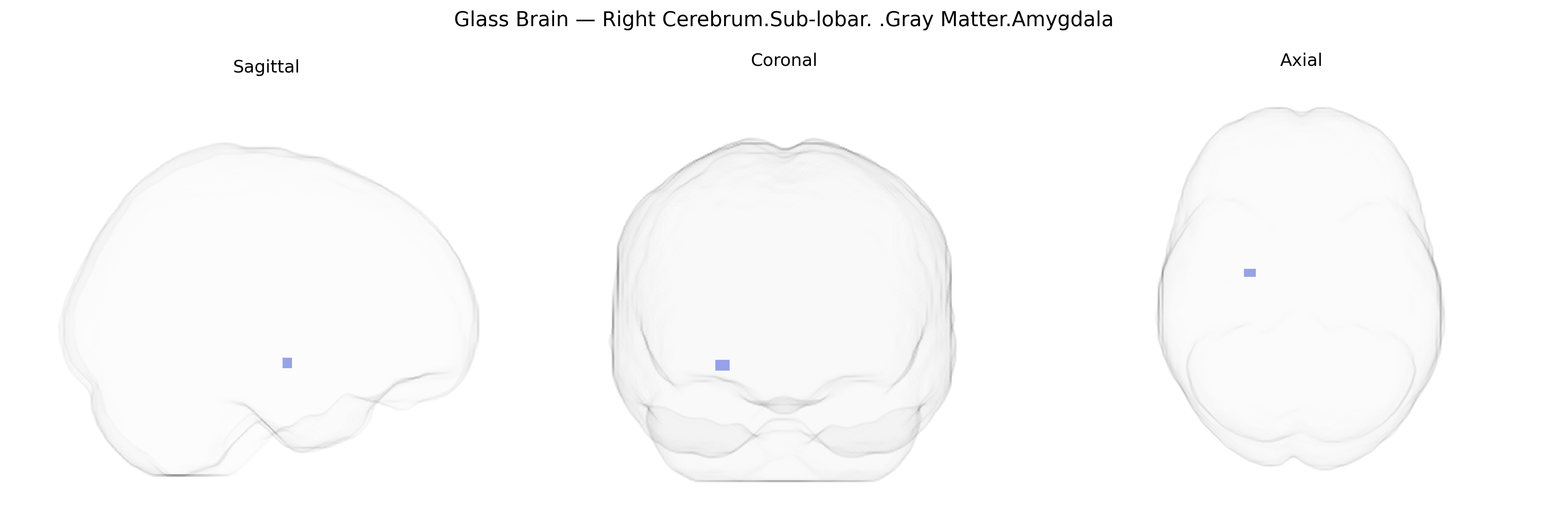
The bilateral Right Cerebrum.Sub-lobar.Gray Matter.Amygdala, as defined in the Talairach 2 mm Atlas, corresponds to the right amygdaloid complex located within the medial temporal lobe, deep to the uncus and anterior to the hippocampus. This sub-lobar gray matter structure is composed of multiple nuclei (including basolateral, corticomedial, and central groups) that integrate sensory, contextual, and interoceptive information to modulate emotional processing, particularly fear and threat detection, as well as aspects of reward, social cognition, and affective memory. The right amygdala, in many studies, has been implicated in rapid detection and automatic evaluation of emotionally salient stimuli, autonomic and endocrine responses to stress, and coordination with prefrontal and hippocampal regions for emotional learning and consolidation of emotional memories.
The bilateral right sub-lobar gray matter amygdala region, as defined in the Talairach 2 mm atlas, has been repeatedly implicated in genetic studies linking structure and function of the amygdala to psychiatric and behavioral traits. GWAS and imaging-genetics work show that common variants in genes related to synaptic plasticity and neurotransmission—such as BDNF (e.g., Val66Met), SLC6A4 (serotonin transporter), COMT, and various glutamatergic and GABAergic genes—are associated with amygdala volume, reactivity to emotional stimuli, and connectivity with prefrontal cortex. Large-scale GWAS of subcortical brain volumes (e.g., ENIGMA consortium studies) have identified multiple loci influencing amygdala size, including variants near genes involved in neurodevelopment, cell adhesion, and axonal guidance, though individual effects are small and highly polygenic. Genetic variation affecting right amygdala structure and function has been linked to risk or endophenotypes for disorders such as major depressive disorder, anxiety disorders (including social anxiety and generalized anxiety), post-traumatic stress disorder, schizophrenia, bipolar disorder, and autism spectrum disorder, as well as traits like neuroticism, aggression, stress reactivity, and emotion recognition. Polygenic risk scores for several psychiatric conditions correlate with alterations in amygdala volume and activation, indicating shared genetic architecture between this region and vulnerability to psychopathology, although specific associations are generally not hemisphere- or sub-lobar–exclusive and reflect distributed brain–gene networks rather than a single locus.
Overview generated by GPT-4o (2026).
Region ID: 358
Hemisphere: bilateral
Atlas: Talairach labels 2mm
Full Quality Version: Download MP4
Full Quality Version: Download MP4
Wali Sidiqyar*, Gaurav Rudravaram*, Elyssa M. McMaster, Trent M. Schwartz, Adam M. Saunders, Kurt G. Schilling, Bennett A. Landman "Introducing SPINS: A Shared Public Visualization Library of Neuroanatomical Structures." Medical Imaging with Deep Learning- short paper
This resource is licensed under CC0 1.0 Universal (Public Domain).