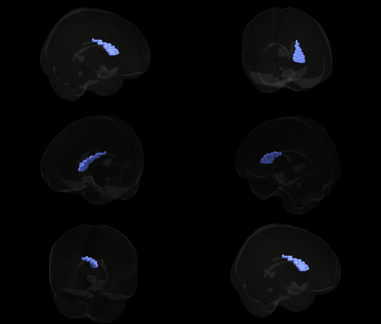
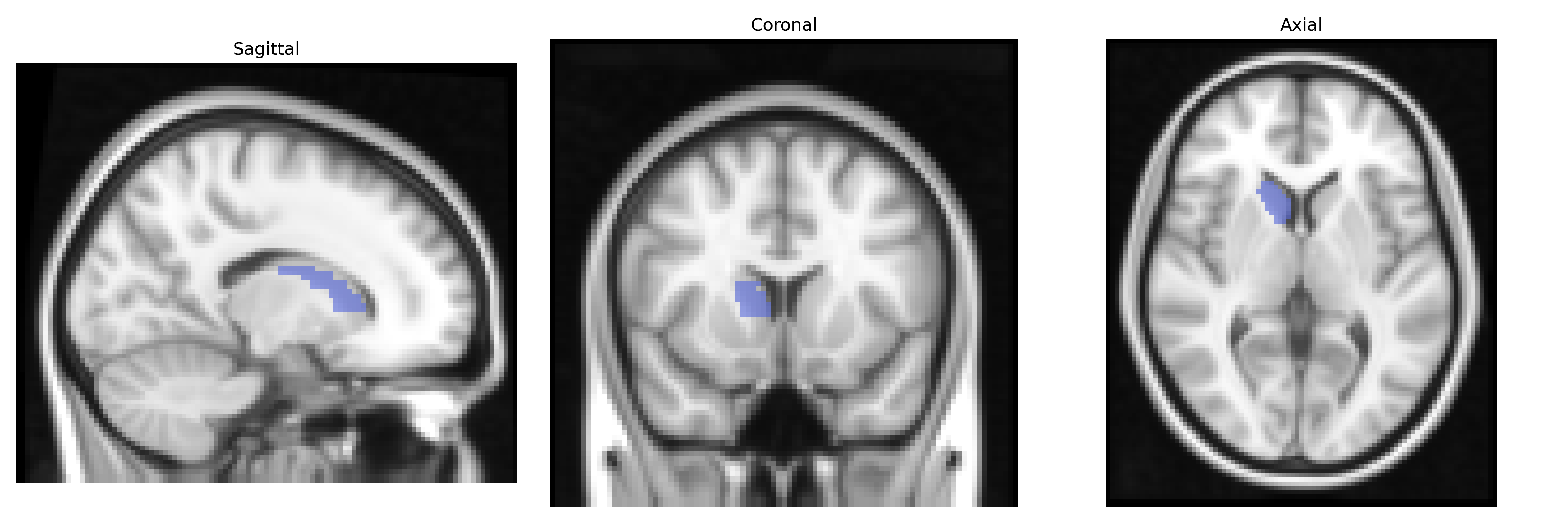
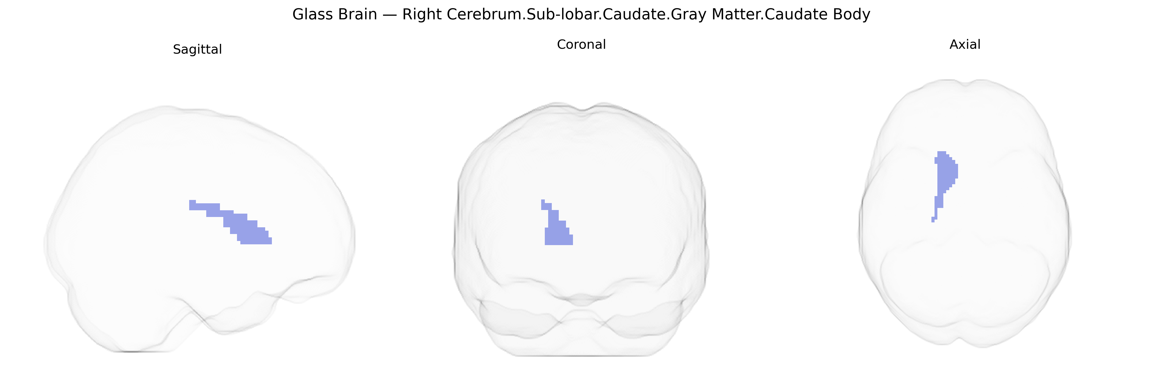
The bilateral Right Cerebrum.Sub-lobar.Caudate.Gray Matter.Caudate Body corresponds to the main portion of the caudate nucleus, a C-shaped gray matter structure within the basal ganglia of the sub-lobar region of the cerebral hemispheres. The caudate body is critically involved in motor control, action selection, procedural learning, and various aspects of cognition, including executive functions and reward-related processing. It receives extensive glutamatergic input from the cortex and dopaminergic input from the substantia nigra, integrating these signals to modulate output to other basal ganglia structures via GABAergic projections. Functionally, the caudate body contributes to the regulation of movement, habit formation, and goal-directed behaviors, and its dysfunction has been implicated in neurological and psychiatric conditions such as Parkinson’s disease, Huntington’s disease, obsessive–compulsive disorder, and attention-deficit/hyperactivity disorder.
Caudate nucleus
The bilateral right caudate body, a sub-lobar gray matter structure within the cerebrum, has been repeatedly implicated in genetic studies of brain morphology, cognition, and neuropsychiatric disease. GWAS of subcortical volumes (e.g., ENIGMA and UK Biobank cohorts) have identified multiple loci influencing caudate volume, including variants near/in genes such as MIR137, DCC, FAT3, KTN1, HMGA2, and FAF1, among others, many of which are involved in neurodevelopment, axon guidance, and synaptic function. Polygenic risk scores for schizophrenia, bipolar disorder, major depressive disorder, ADHD, and autism spectrum disorder show associations with caudate structure, consistent with the caudate’s role in frontostriatal circuits underlying motivation, reward, and executive control. Huntington’s disease, caused by CAG repeat expansion in the HTT gene, features early and prominent caudate atrophy, while risk variants for Parkinson’s disease and related movement disorders have been linked to basal ganglia circuitry that includes the caudate. Imaging genetics studies also connect caudate morphology and activation with allelic variation in dopaminergic genes (e.g., DRD2, DRD3, COMT) and glutamatergic or synaptic genes, relating these to traits such as cognitive performance, impulsivity, and substance use. Although most GWAS do not distinguish Talairach-defined subregions like “Caudate Body” in a fine-grained way, the identified genetic influences on caudate volume and function broadly apply to this region and support its role as a key genetically modulated node in neuropsychiatric and cognitive phenotypes.
Overview generated by GPT-4o (2026).
Region ID: 693
Hemisphere: bilateral
Atlas: Talairach labels 2mm
Full Quality Version: Download MP4
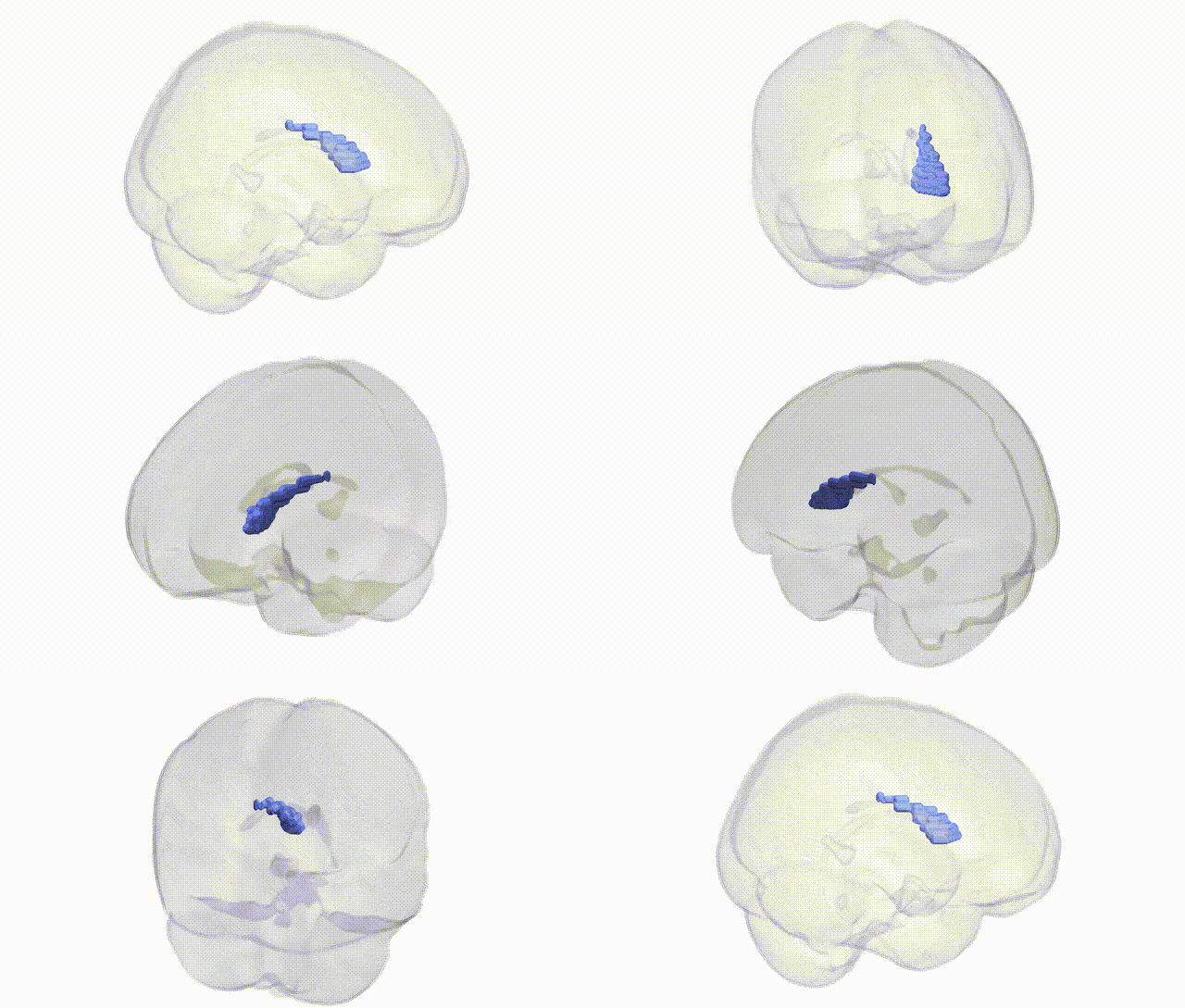
Full Quality Version: Download MP4
Wali Sidiqyar*, Gaurav Rudravaram*, Elyssa M. McMaster, Trent M. Schwartz, Adam M. Saunders, Kurt G. Schilling, Bennett A. Landman "Introducing SPINS: A Shared Public Visualization Library of Neuroanatomical Structures." Medical Imaging with Deep Learning- short paper
This resource is licensed under CC0 1.0 Universal (Public Domain).