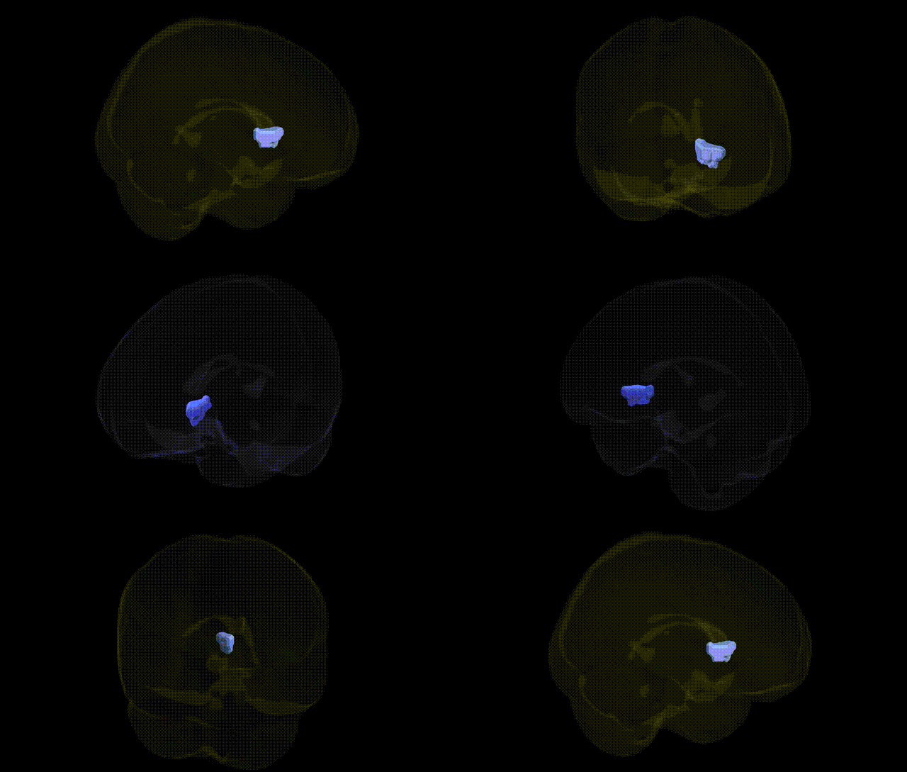
The bilateral Right Cerebrum.Sub-lobar.Caudate.Gray Matter.Caudate Head corresponds to the anterior portion of the caudate nucleus, a major component of the dorsal striatum within the basal ganglia circuitry. This gray matter region is heavily interconnected with prefrontal and limbic cortices via cortico-striato-thalamo-cortical loops, and is critically involved in higher-order executive functions, action selection, goal-directed behavior, and aspects of reward-based learning. The caudate head receives dense dopaminergic input from the substantia nigra pars compacta and participates in modulation of motor output as well as in cognitive and emotional processes; dysfunction in this region has been implicated in movement disorders (e.g., Parkinson’s disease, Huntington’s disease) and neuropsychiatric conditions such as obsessive–compulsive disorder and attention-deficit/hyperactivity disorder. There is no direct Wikipedia article for “Caudate Head”; a closely related structure is the Caudate nucleus.
The caudate head, a sub-lobar gray matter structure in the right cerebrum, has been repeatedly implicated in genetic studies of brain morphology and neuropsychiatric traits. GWAS of subcortical volumes from large cohorts (e.g., ENIGMA, UK Biobank) have identified common variants near genes such as HMGA2, IGF1, FAT3, DRD2, and KTN1 that influence caudate volume and shape, with several loci showing bilateral effects on the caudate head. These genetic influences are often pleiotropic, overlapping with variants associated with general cognitive ability, educational attainment, and intelligence, consistent with the caudate head’s role in frontostriatal circuits supporting executive function, learning, and motivation. Psychiatric and neurodevelopmental GWAS have linked caudate-related genetics to disorders including schizophrenia, ADHD, major depressive disorder, obsessive–compulsive disorder, and Tourette’s syndrome, in part through dopaminergic signaling genes and synaptic plasticity pathways that alter striatal structure or function. Additionally, polygenic risk scores for Parkinson’s disease, Huntington’s disease (driven by HTT CAG repeat expansion), and other movement disorders are associated with caudate head atrophy or altered morphology, reflecting the vulnerability of this region to genetic disruptions in basal ganglia circuitry. Overall, the bilateral caudate head emerges as a genetically influenced hub in dopaminergic and corticostriatal networks, with its volume and integrity shaped by multiple loci that also contribute to psychiatric disease risk, cognitive traits, and motor control.
Overview generated by GPT-4o (2026).
Region ID: 465
Hemisphere: bilateral
Atlas: Talairach labels 2mm
Full Quality Version: Download MP4
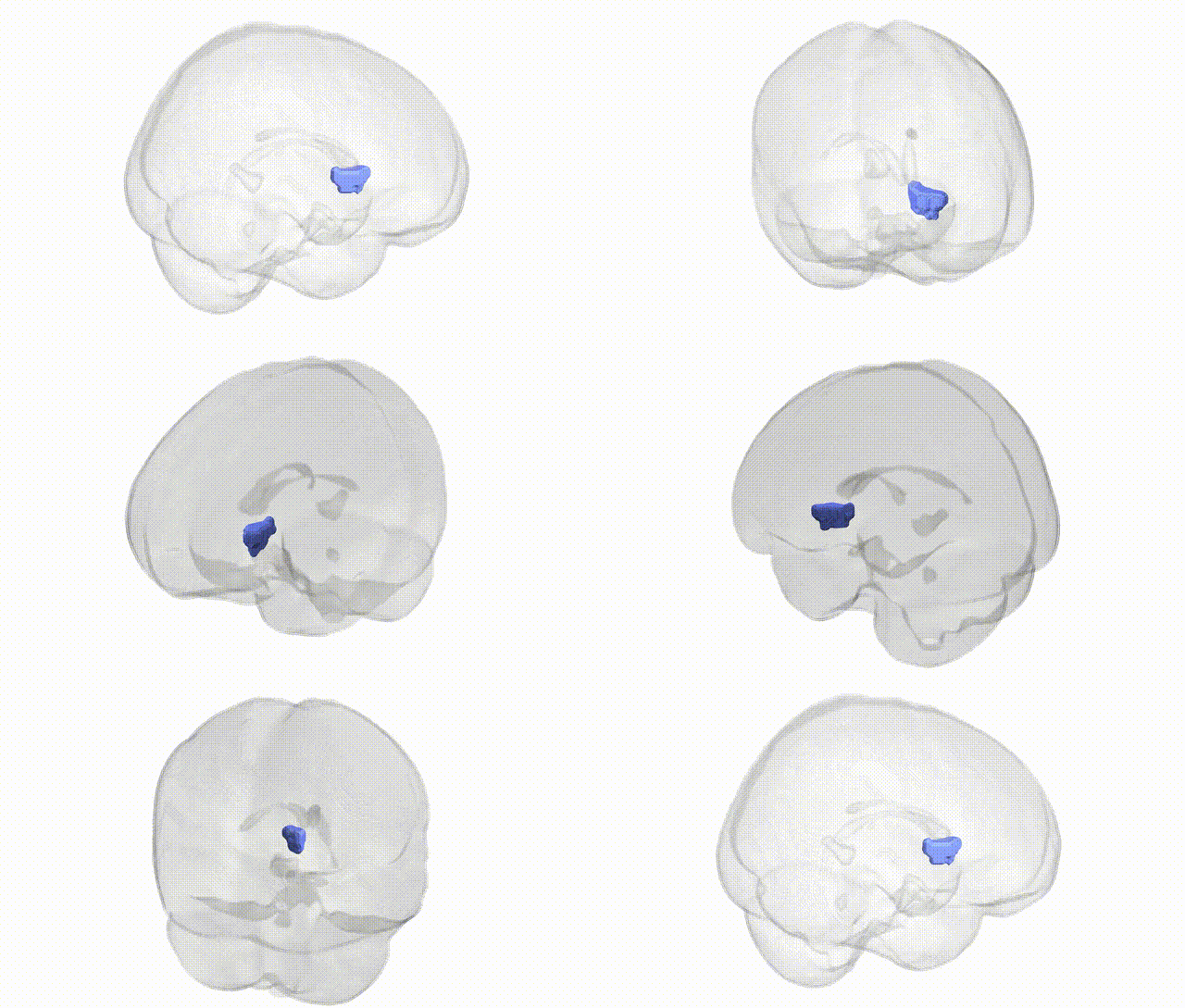
Full Quality Version: Download MP4
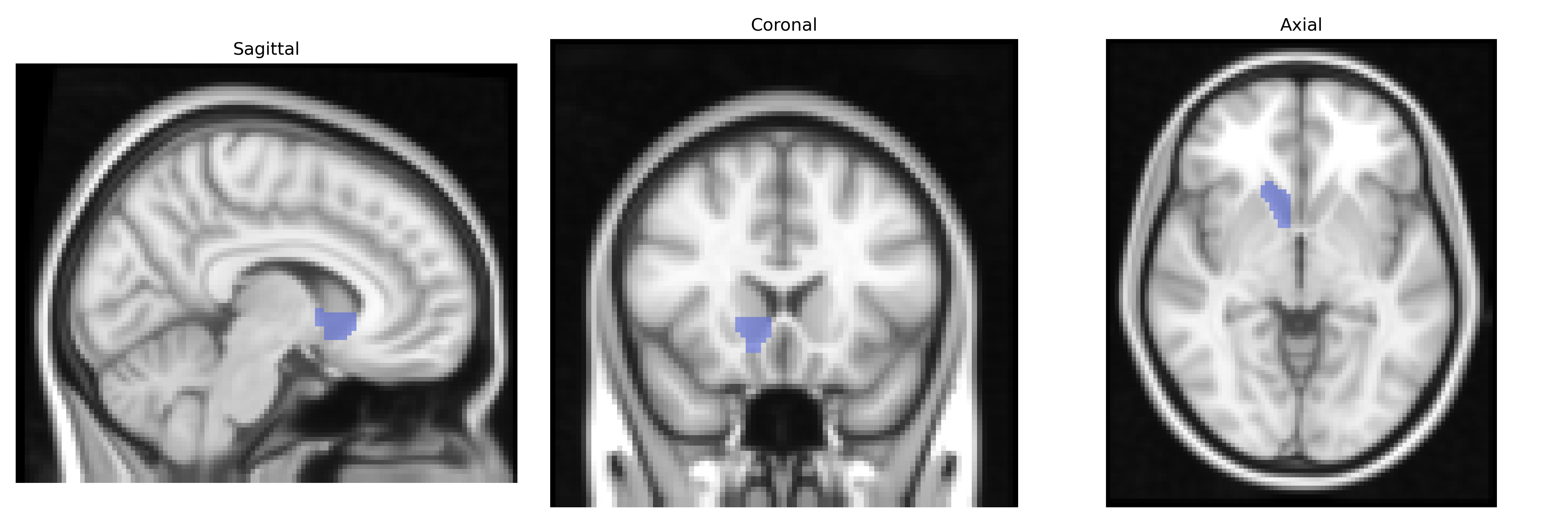
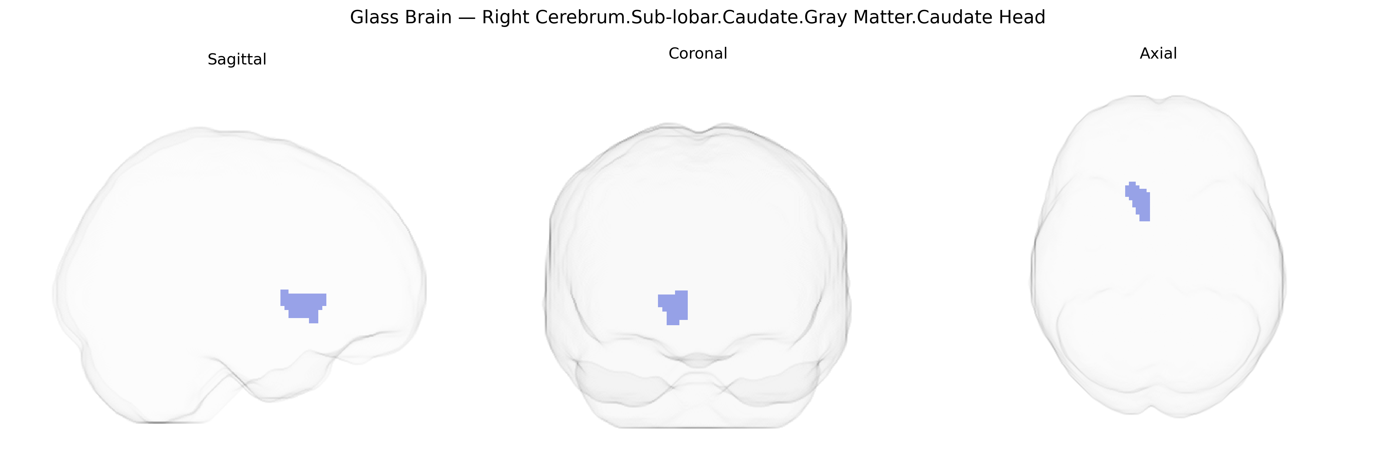
Wali Sidiqyar*, Gaurav Rudravaram*, Elyssa M. McMaster, Trent M. Schwartz, Adam M. Saunders, Kurt G. Schilling, Bennett A. Landman "Introducing SPINS: A Shared Public Visualization Library of Neuroanatomical Structures." Medical Imaging with Deep Learning- short paper
This resource is licensed under CC0 1.0 Universal (Public Domain).