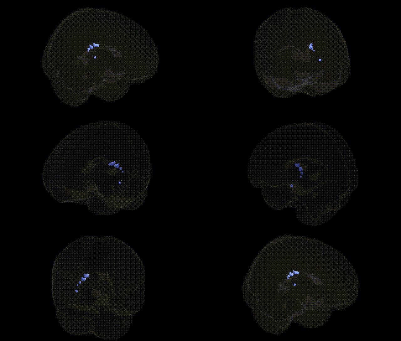
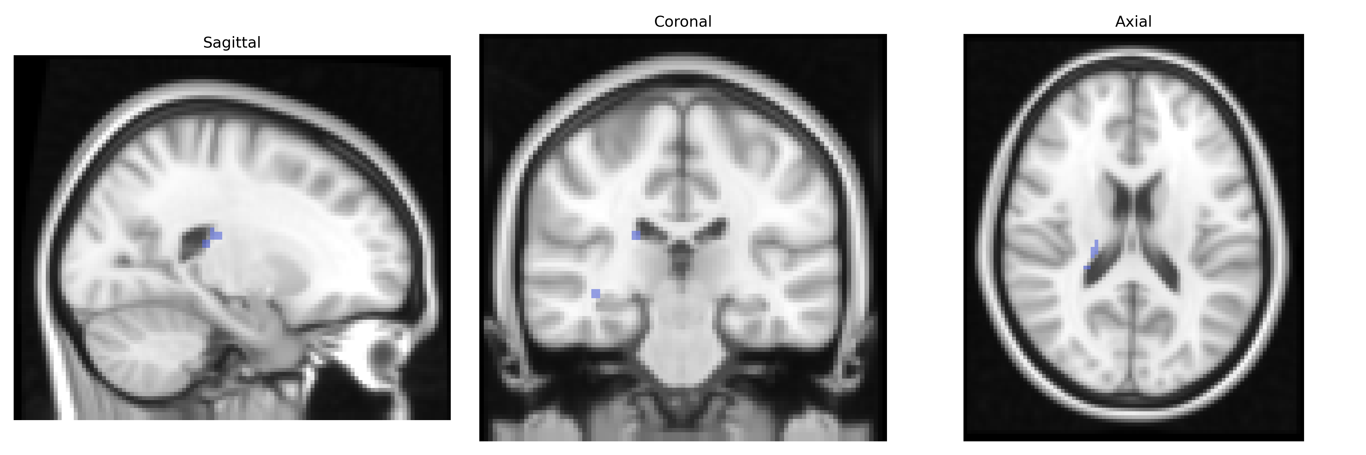
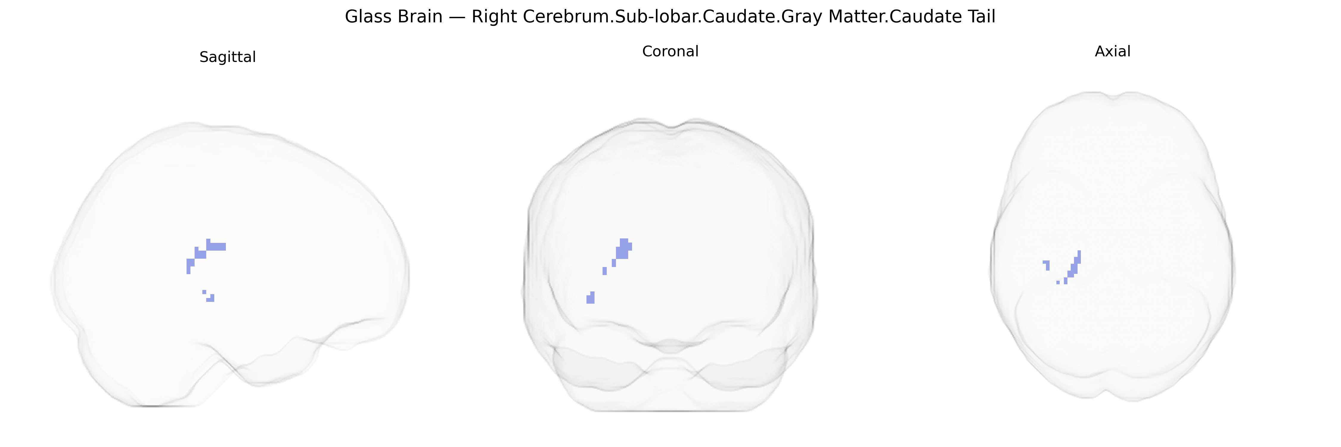
The bilateral Right Cerebrum.Sub-lobar.Caudate.Gray Matter.Caudate Tail corresponds to the posterior portion of the caudate nucleus within the subcortical gray matter of the right hemisphere, as defined in the Talairach 2 mm Atlas. The caudate tail is a curved, elongated segment that follows the lateral ventricle into the temporal lobe, forming part of the dorsal striatum and integrating inputs from associative and limbic cortical areas. Functionally, it contributes to motor control, procedural and habit learning, and higher-order cognitive processes, including reward-based learning and action selection. It receives dopaminergic input from the midbrain and projects via basal ganglia circuits to thalamic and cortical regions, playing a role in the modulation of behavior and in disorders such as Parkinson’s disease, Huntington’s disease, and certain psychiatric conditions. There is no direct Wikipedia article for the caudate tail; a closely related structure is the Caudate nucleus.
The caudate tail, part of the dorsal striatum in the sub-lobar gray matter of the right cerebrum, has been implicated in several genetic and neuropsychiatric associations, though usually studied as part of the broader caudate or striatal complex rather than as an isolated tail subregion. Genome-wide association studies of striatal and caudate volume have identified robust associations with variants near genes such as KNTC1, DCC, BDNF, and HRK, among others, reflecting polygenic influences on caudate structure that likely extend to the tail region. Striatal circuitry, including the caudate tail, is strongly modulated by dopaminergic and glutamatergic pathways, and genes involved in these systems (e.g., DRD2, DRD3, DAT1/SLC6A3, COMT, GRIN2B) have been repeatedly linked to traits involving reward learning, habit formation, and reinforcement processing, functions to which the caudate tail contributes. Structural and functional imaging-genetics studies have associated caudate morphology and activation with neurodevelopmental and psychiatric disorders such as Huntington’s disease (HTT expansions with marked caudate atrophy), obsessive-compulsive disorder (polygenic risk involving synaptic and neurotransmission genes and altered caudate/striatal structure), Tourette syndrome (variants in CNTNAP2, SLITRK1 and related synaptic genes impacting cortico-striatal circuits), attention-deficit/hyperactivity disorder (risk alleles in dopaminergic and synaptic genes affecting fronto-striatal connectivity), and schizophrenia (polygenic risk scores linked to caudate and broader striatal volume and activation changes). Additional GWAS and imaging-genetics work has connected caudate and striatal measures to traits such as general cognitive ability, educational attainment, smoking and substance use, and body mass index, with loci in neurodevelopmental, synaptic, and transcriptional regulation genes influencing these phenotypes. While Talairach-specific “caudate tail” labels are not typically the direct focus of GWAS, current evidence indicates that this region shares the broader striatal genetic architecture shaping reward-related learning, habit circuitry, and vulnerability to several psychiatric and behavioral traits.
Overview generated by GPT-4o (2026).
Region ID: 436
Hemisphere: bilateral
Atlas: Talairach labels 2mm
Full Quality Version: Download MP4
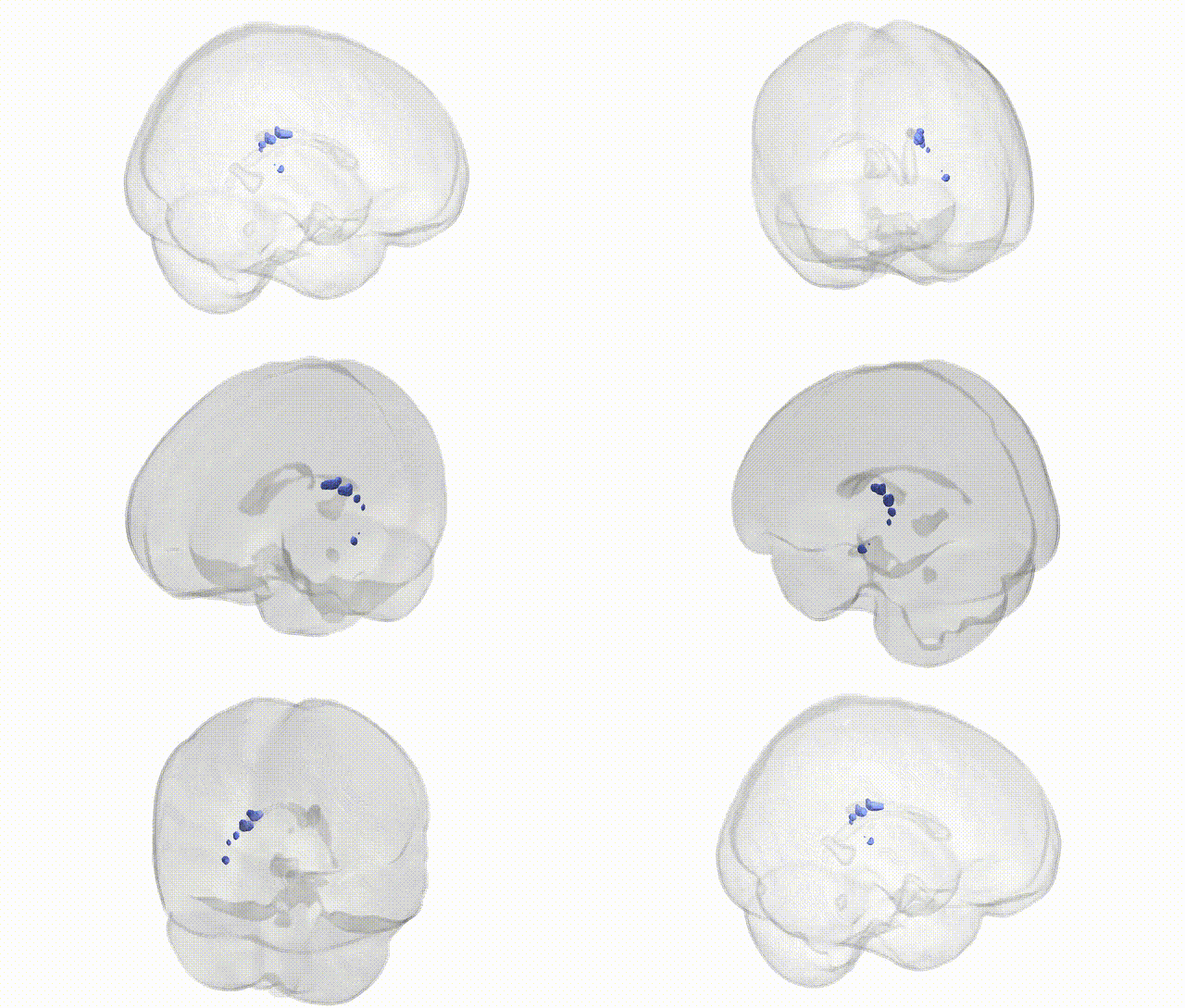
Full Quality Version: Download MP4
Wali Sidiqyar*, Gaurav Rudravaram*, Elyssa M. McMaster, Trent M. Schwartz, Adam M. Saunders, Kurt G. Schilling, Bennett A. Landman "Introducing SPINS: A Shared Public Visualization Library of Neuroanatomical Structures." Medical Imaging with Deep Learning- short paper
This resource is licensed under CC0 1.0 Universal (Public Domain).