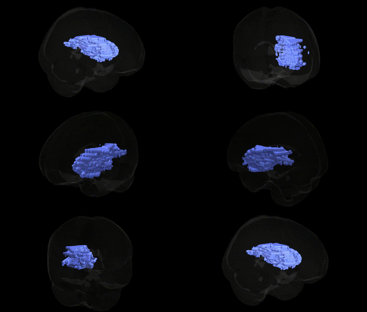
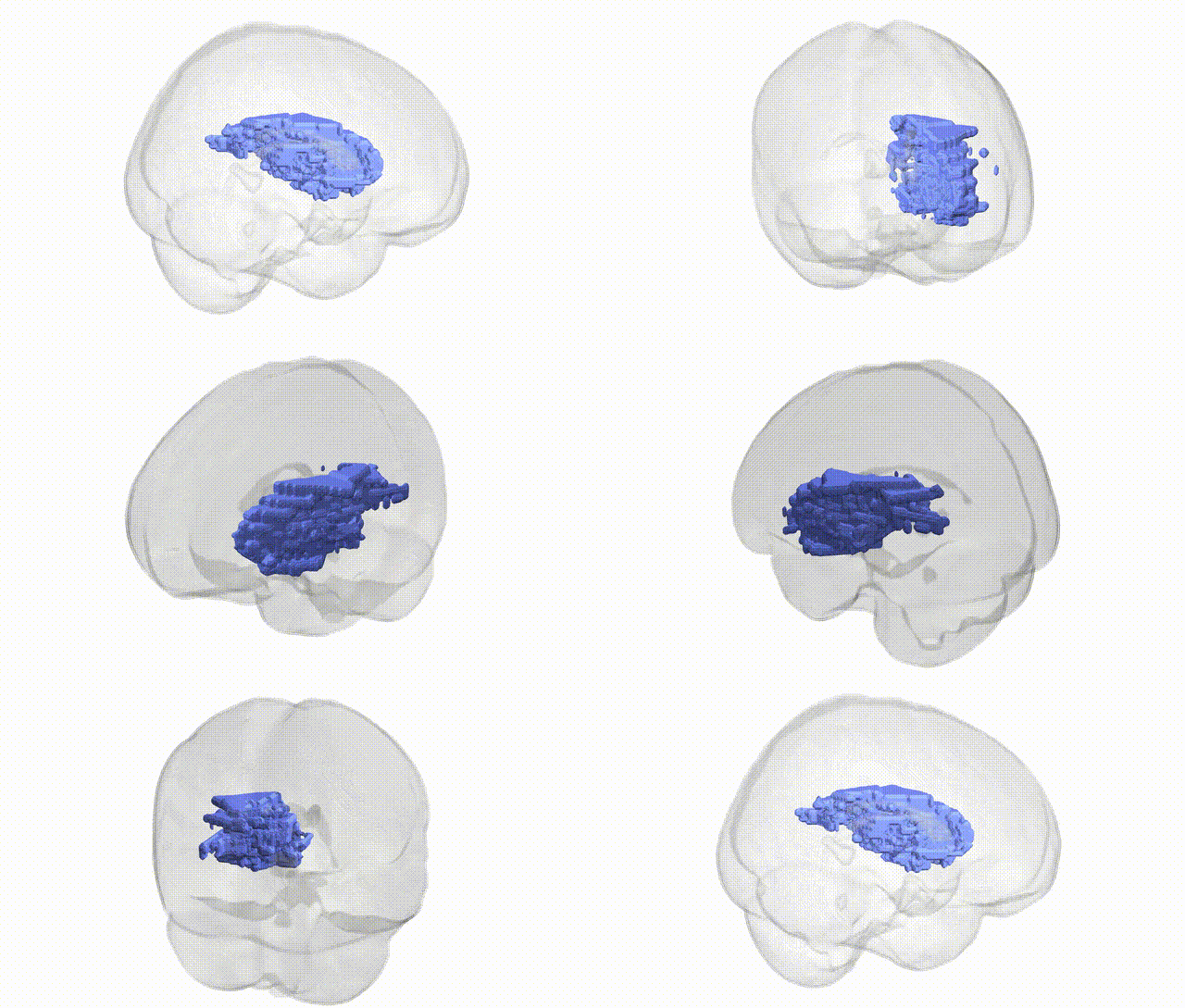
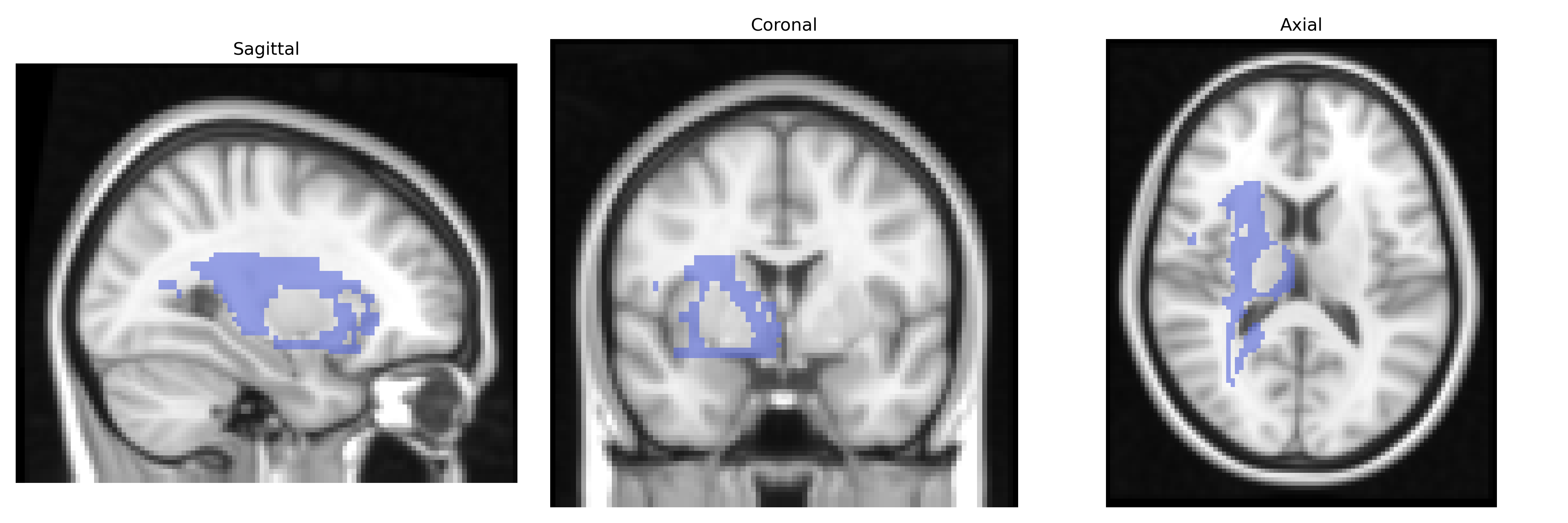
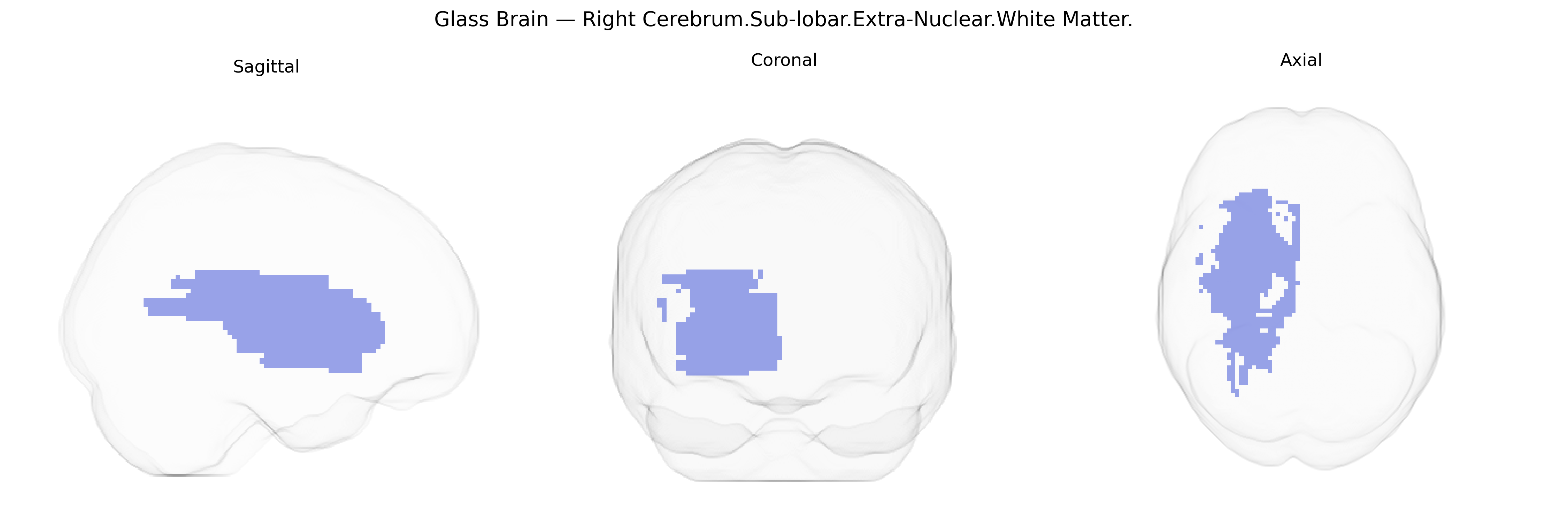
The bilateral Right Cerebrum.Sub-lobar.Extra-Nuclear.White Matter refers to the deep, subcortical white matter tracts located lateral to and surrounding the basal ganglia and thalamus within the right cerebral hemisphere. These regions consist predominantly of myelinated axons forming portions of major projection, association, and commissural pathways, including segments of the internal capsule and adjacent periventricular and subcortical fiber systems. Functionally, this extra-nuclear white matter supports long-range communication between cortical and subcortical structures, contributing to motor control, sensory integration, cognition, and language processes by enabling rapid signal conduction across distributed neural networks. There is no direct Wikipedia article for this specific Talairach label; a closely related structure is the White matter.
Genetic associations specific to the bilateral Right Cerebrum.Sub-lobar.Extra-Nuclear.White Matter region, as defined by the Talairach 2 mm atlas, are not well characterized at the fine-grained label level, but numerous imaging genetics and GWAS studies implicate white-matter microstructure and volume in this general sub-lobar extra-nuclear territory. Common variants in genes involved in axonal guidance, myelination, and neurodevelopment—such as CNTNAP2, NRG1, MAG, LINGO1, and those in the oligodendrocyte lineage pathways—have been associated with variation in diffusion MRI metrics (e.g., fractional anisotropy) and white-matter integrity across frontal and sub-lobar regions, including right-hemisphere tracts. Large-scale GWAS of brain imaging phenotypes (e.g., UK Biobank, ENIGMA) have identified loci near genes such as BDNF, VAC14, DCC, and FOXP2 that modulate global and regional white-matter structure and connectivity, which in turn show genetic correlations with neuropsychiatric disorders (schizophrenia, bipolar disorder, major depression, ADHD, and autism spectrum disorder), cognitive traits (intelligence, processing speed, working memory), and risk behaviors. White-matter abnormalities in right sub-lobar regions are consistently reported in genetic studies of schizophrenia and bipolar disorder (often linked to polygenic risk scores), and GWAS of traumatic brain injury, migraine, and small-vessel disease suggest that susceptibility alleles influencing vascular integrity and inflammatory processes may also affect white-matter health in these territories. However, current evidence maps primarily to tracts and lobar/sub-lobar white matter more broadly rather than to the specific “Right Cerebrum.Sub-lobar.Extra-Nuclear.White Matter” label, so associations should be interpreted as regionally overlapping rather than anatomically precise.
Overview generated by GPT-4o (2026).
Region ID: 348
Hemisphere: bilateral
Atlas: Talairach labels 2mm
Full Quality Version: Download MP4
Full Quality Version: Download MP4
Wali Sidiqyar*, Gaurav Rudravaram*, Elyssa M. McMaster, Trent M. Schwartz, Adam M. Saunders, Kurt G. Schilling, Bennett A. Landman "Introducing SPINS: A Shared Public Visualization Library of Neuroanatomical Structures." Medical Imaging with Deep Learning- short paper
This resource is licensed under CC0 1.0 Universal (Public Domain).