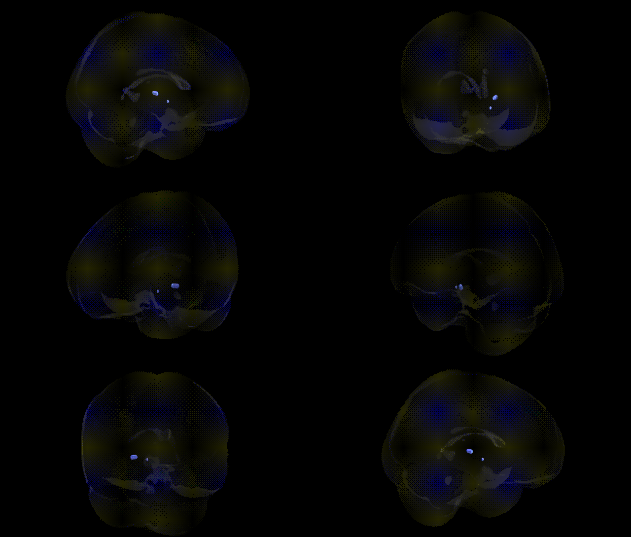
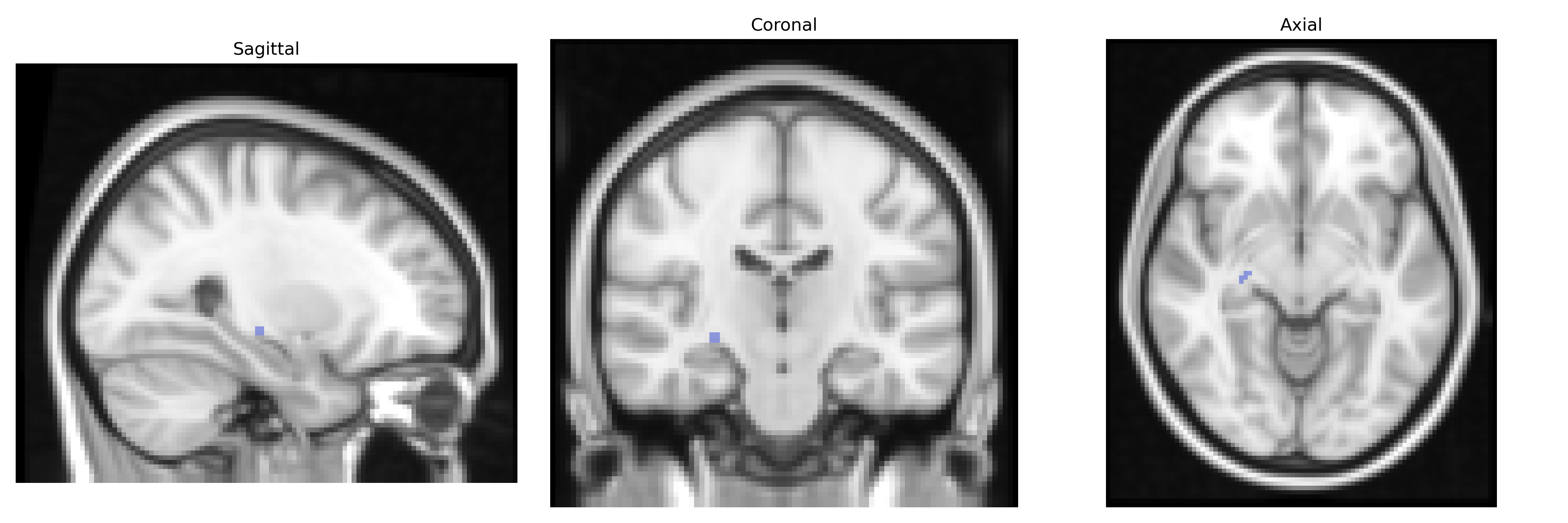
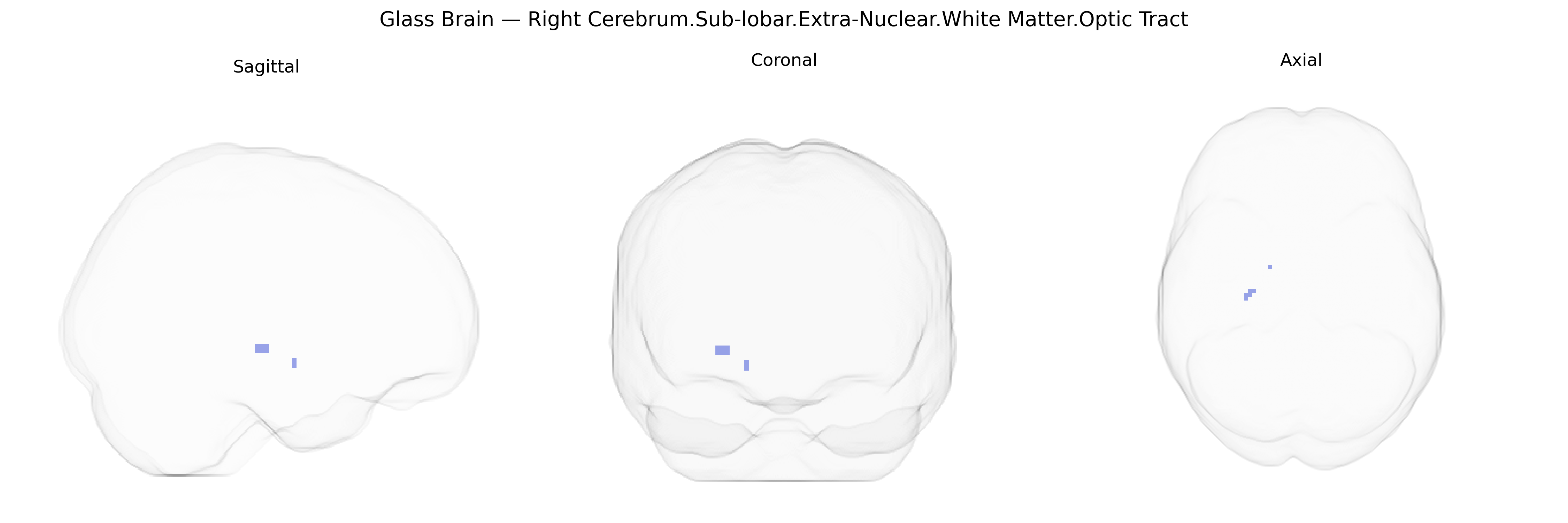
The bilateral Right Cerebrum.Sub-lobar.Extra-Nuclear.White Matter.Optic Tract region in the Talairach 2 mm Atlas corresponds to the right-sided segment of the optic tract, a myelinated white matter bundle that carries visual information from the retinal ganglion cells in both eyes to subcortical visual relay centers. After leaving the optic chiasm, fibers in the optic tract primarily project to the lateral geniculate nucleus of the thalamus, with collateral branches to the superior colliculus, pretectal nuclei, and other visual reflex centers, supporting conscious visual perception, eye movement control, pupillary light reflexes, and visuospatial orienting. Functionally, this tract is critical for transmitting retinotopically organized signals that preserve visual field mapping, and lesions in this region typically produce characteristic visual field defects such as homonymous hemianopia. There is no direct link for this specific bilateral right Talairach label; a related structure is the Optic tract.
Genetic associations specifically targeting the bilateral Right Cerebrum.Sub-lobar.Extra-Nuclear.White Matter.Optic Tract region in the Talairach 2 mm atlas are sparse, but broader imaging-genetics and GWAS work implicates several loci and pathways in optic tract and visual white‑matter microstructure. Large neuroimaging GWAS (e.g., UK Biobank–based) have shown heritable variation in diffusion tensor imaging measures (such as fractional anisotropy and mean diffusivity) within visual pathway tracts, with significant associations to genes involved in axonal guidance, myelination, and neurodevelopment, including variants near genes such as CNTN4, ROBO1/2, and MAG that influence white‑matter integrity and visual system architecture. Multiple sclerosis and other demyelinating disorders, which often affect the optic tract, have robust GWAS signals in immune‑related genes (HLA-DRB1, IL2RA, TNFRSF1A) whose pathogenic mechanisms include inflammatory damage to central visual white matter. Inherited optic neuropathies (notably Leber hereditary optic neuropathy, associated with mitochondrial DNA mutations such as MT-ND1, MT-ND4, and MT-ND6) and some nuclear gene–related optic atrophies (e.g., OPA1, MFN2) involve degeneration along the optic nerve and tract, and imaging studies frequently demonstrate corresponding structural changes in this region. More broadly, polygenic influences on visual acuity, refractive error, and glaucoma risk (e.g., variants near MYOC, CAV1/CAV2, SIX6) have been linked to alterations in optic nerve and tract structure, suggesting that visual system GWAS signals converge on the development, maintenance, and vulnerability of the optic tract white matter, even if most studies report tract-based metrics rather than Talairach-defined regional labels.
Overview generated by GPT-4o (2026).
Region ID: 361
Hemisphere: bilateral
Atlas: Talairach labels 2mm
Full Quality Version: Download MP4
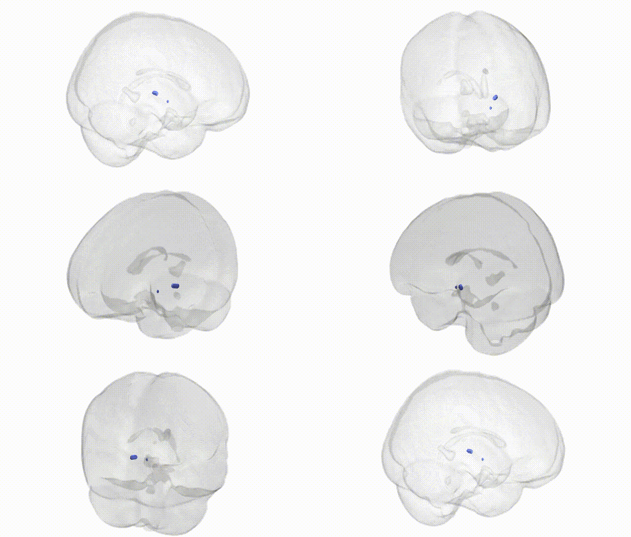
Full Quality Version: Download MP4
Wali Sidiqyar*, Gaurav Rudravaram*, Elyssa M. McMaster, Trent M. Schwartz, Adam M. Saunders, Kurt G. Schilling, Bennett A. Landman "Introducing SPINS: A Shared Public Visualization Library of Neuroanatomical Structures." Medical Imaging with Deep Learning- short paper
This resource is licensed under CC0 1.0 Universal (Public Domain).