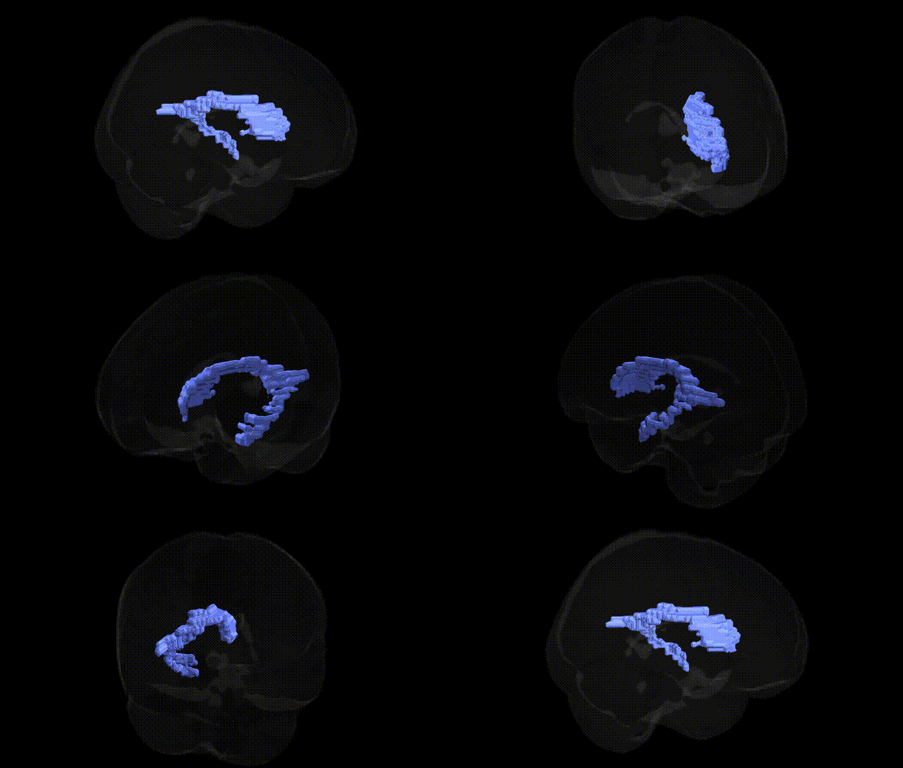
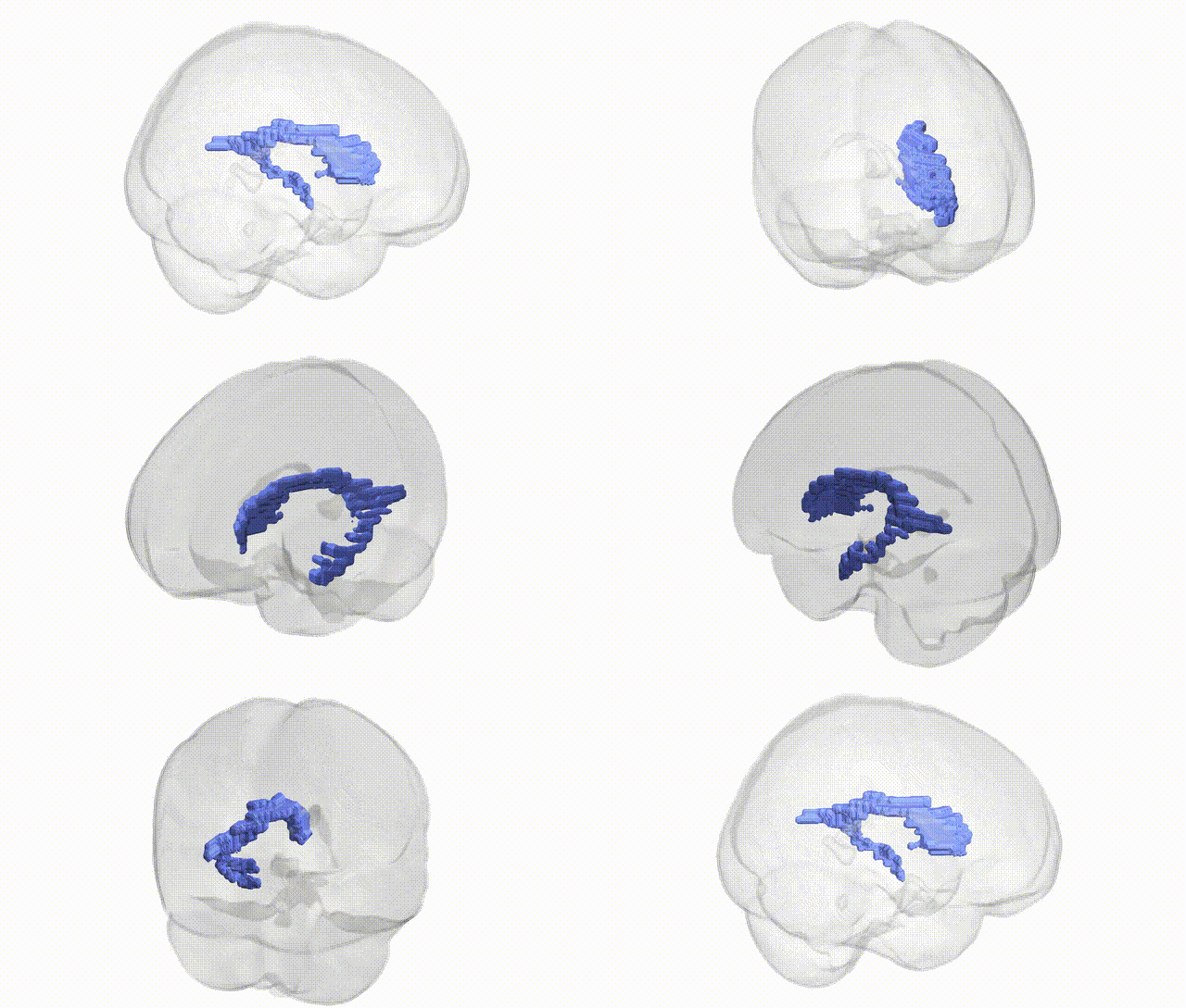
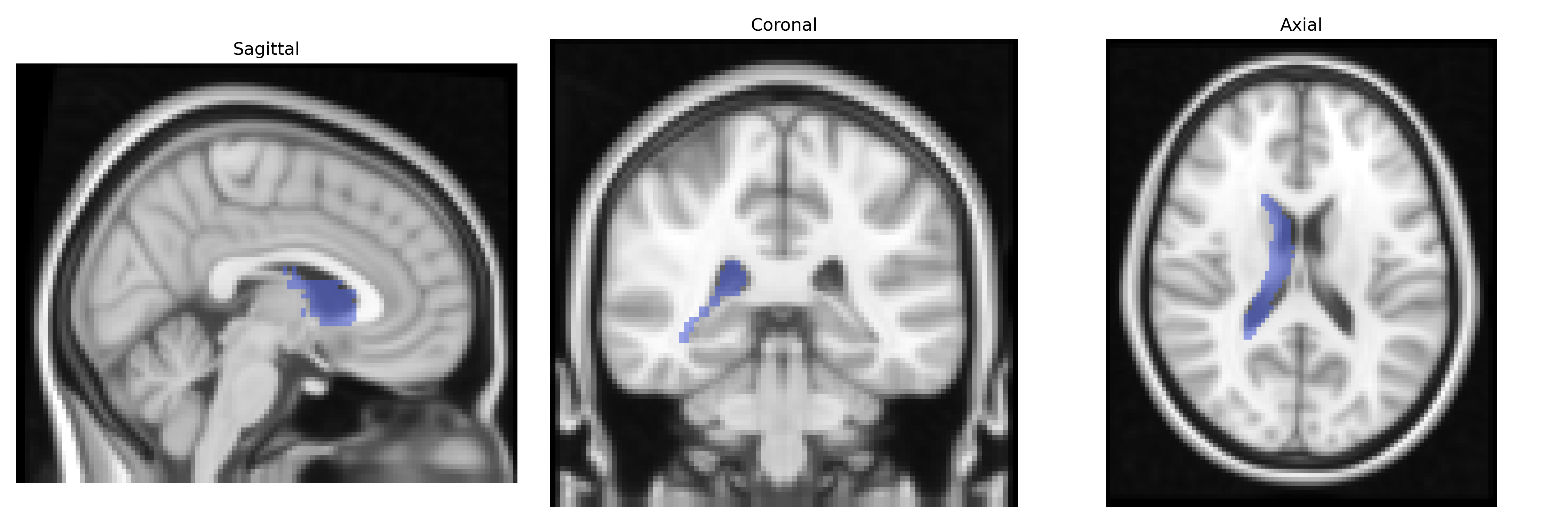
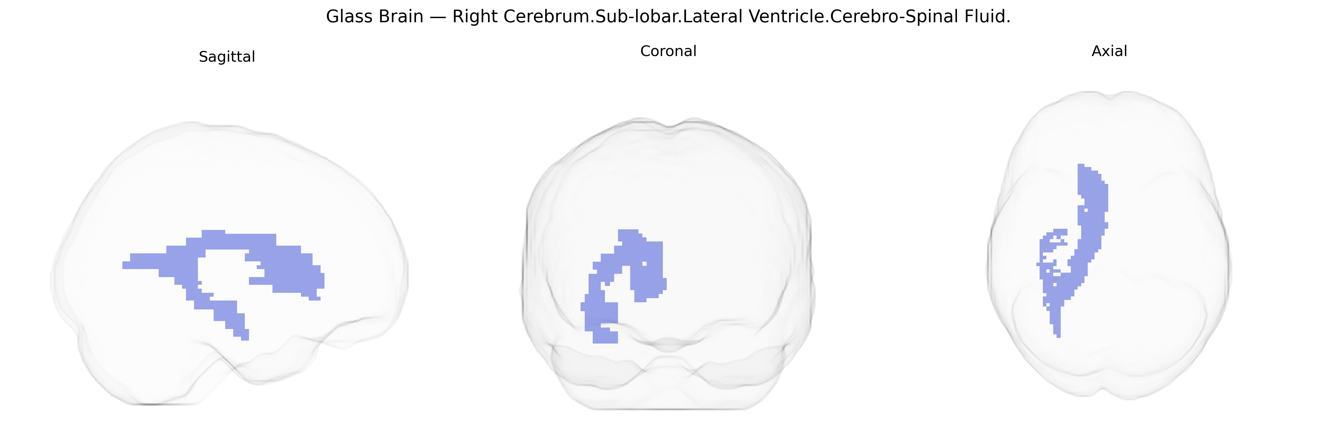
The bilateral Right Cerebrum.Sub-lobar.Lateral Ventricle.Cerebro-Spinal Fluid region, as defined in the Talairach 2 mm Atlas, corresponds to cerebrospinal fluid (CSF) occupying the right lateral ventricle within the sub-lobar territory of the cerebrum. The lateral ventricle is a paired, C-shaped cavity situated deep within the cerebral hemispheres, extending through the frontal, parietal, temporal, and occipital lobes. It is lined by ependymal cells and communicates with the third ventricle via the interventricular foramen (foramen of Monro). CSF within this ventricular space is produced predominantly by the choroid plexus and plays essential roles in cushioning neural tissue, maintaining intracranial pressure, and facilitating the transport of nutrients and waste products. In neuroimaging and stereotaxic frameworks like Talairach, this label denotes fluid-filled voxel locations rather than gray or white matter structures. There is no specific Wikipedia article for “Right Cerebrum.Sub-lobar.Lateral Ventricle.Cerebro-Spinal Fluid”; a closely related structure is the Lateral ventricle.
Genetic associations involving the bilateral right sub-lobar lateral ventricle cerebrospinal fluid region, as defined in the Talairach 2 mm atlas, primarily derive from GWAS of brain structure and ventricular volume rather than region-specific functional studies. Multiple large-scale imaging genetics consortia (e.g., ENIGMA, UK Biobank) have identified loci near genes such as NRXN1, PLEKHM1, MAPT, SLC39A8, and others associated with global and lateral ventricular volume, which in turn reflect neurodevelopmental and neurodegenerative processes impacting periventricular tissue. Expanded lateral ventricles, indexed by increased CSF volume in these regions, show robust genetic overlap with disorders including schizophrenia, bipolar disorder, major depression, Alzheimer’s disease, and other dementias, as well as traits related to cognitive decline and brain aging. Ventricular enlargement has also been linked through polygenic risk scores and GWAS to genetic architectures influencing brain atrophy, intracranial volume, and white matter microstructure in adjacent regions such as the caudate and corpus callosum. Although few studies isolate “right sub-lobar” CSF specifically, symmetric ventricular measures indicate that common variants affecting neurodevelopment (e.g., synaptic and axonal guidance genes), cerebrovascular integrity, and tau/amyloid pathways contribute to individual differences in CSF-filled ventricular space, making this atlas-defined region a structural readout of genetically influenced brain vulnerability rather than a direct target of gene-specific functional mapping.
Overview generated by GPT-4o (2026).
Region ID: 174
Hemisphere: bilateral
Atlas: Talairach labels 2mm
Full Quality Version: Download MP4
Full Quality Version: Download MP4
Wali Sidiqyar*, Gaurav Rudravaram*, Elyssa M. McMaster, Trent M. Schwartz, Adam M. Saunders, Kurt G. Schilling, Bennett A. Landman "Introducing SPINS: A Shared Public Visualization Library of Neuroanatomical Structures." Medical Imaging with Deep Learning- short paper
This resource is licensed under CC0 1.0 Universal (Public Domain).