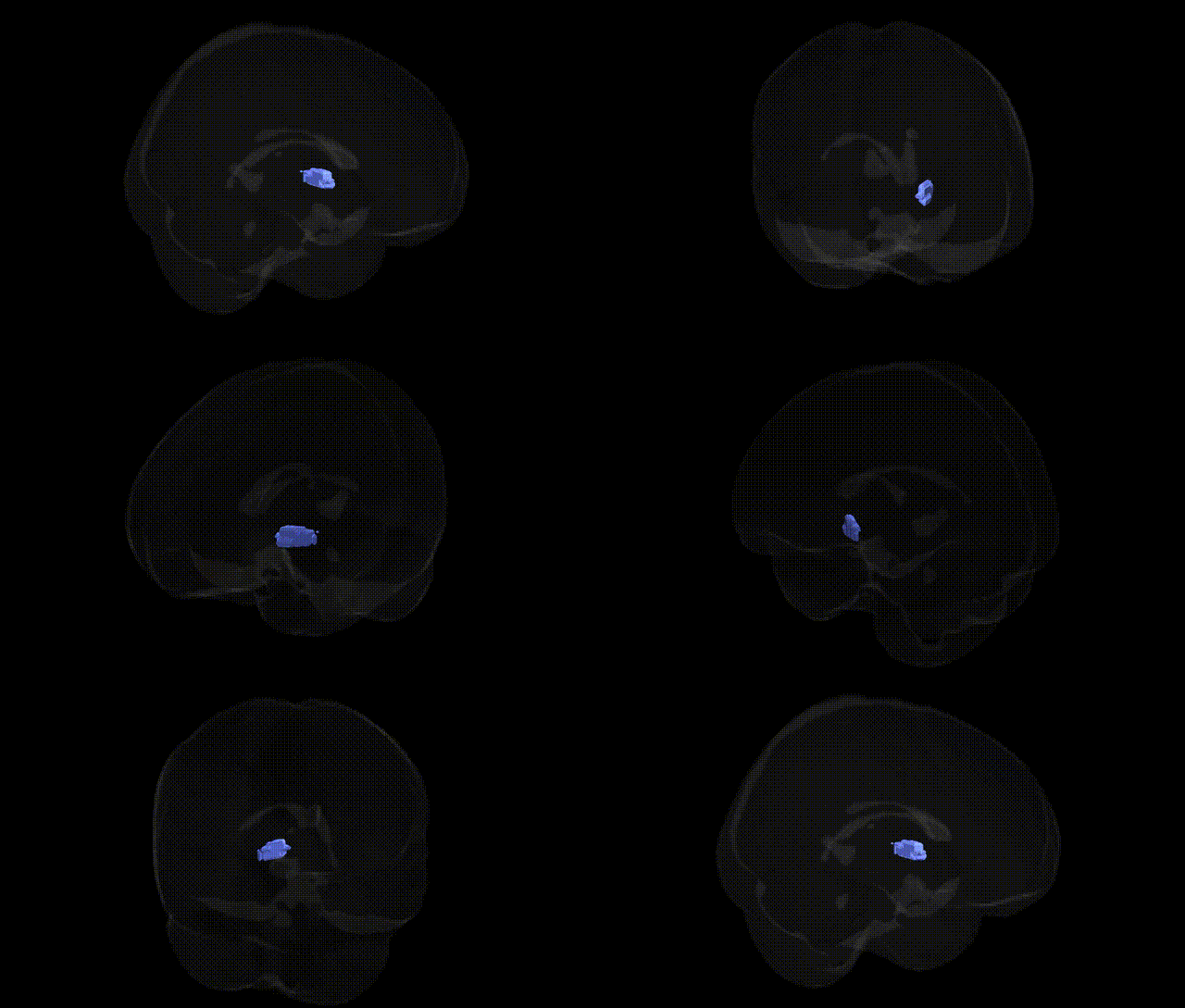
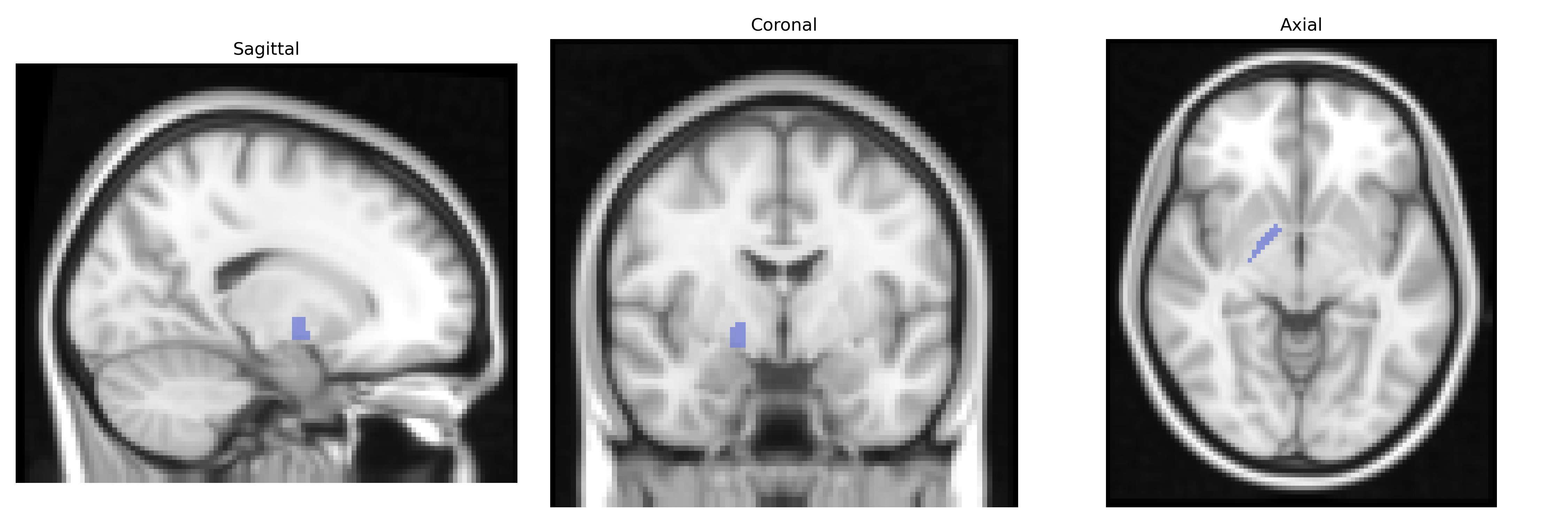
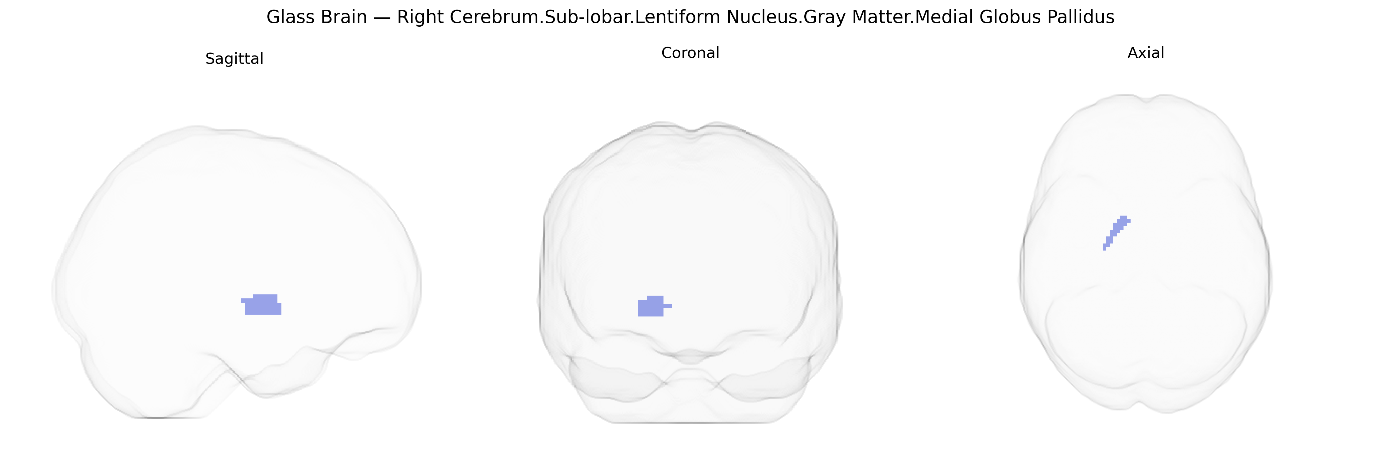
The bilateral Right Cerebrum.Sub-lobar.Lentiform Nucleus.Gray Matter.Medial Globus Pallidus corresponds to the medial segment of the globus pallidus, a major basal ganglia output nucleus situated within the lentiform complex deep to the insular cortex. Composed predominantly of densely packed GABAergic projection neurons oriented in laminar arrangements, this structure receives inhibitory input mainly from the striatum and excitatory modulatory input from the subthalamic nucleus. Its axons form part of the pallidothalamic and pallidosubthalamic pathways, projecting to motor thalamic nuclei and brainstem structures, where it plays a critical role in regulating voluntary movement, muscle tone, and aspects of motor learning via tonic inhibitory control of thalamocortical circuits. Dysfunction of the medial globus pallidus is implicated in movement disorders such as Parkinson’s disease and dystonia, and it is a common target for deep brain stimulation and lesion-based therapies.
Globus pallidus
The medial globus pallidus, part of the lentiform nucleus within the basal ganglia, has been implicated in genetic studies primarily through its role in movement regulation, reward circuitry, and cognitive control. GWAS and imaging-genetics work, including large consortia such as ENIGMA, have identified variants in genes involved in synaptic transmission, neurodevelopment, and neuronal signaling (e.g., DRD2, COMT, BDNF, and glutamatergic and GABAergic pathway genes) that correlate with basal ganglia volume, connectivity, or function, though associations are often reported for the globus pallidus or lentiform nucleus as a whole rather than specifically the medial subdivision. Monogenic disorders affecting pallidal structure and function, such as dystonia (e.g., TOR1A/DYT1, THAP1/DYT6, GNAL, and others), Wilson disease (ATP7B), and some neurodegeneration with brain iron accumulation syndromes (e.g., PANK2, PLA2G6), demonstrate a strong genetic impact on pallidal anatomy and pathology. In psychiatric genetics, risk alleles for schizophrenia, bipolar disorder, major depression, and obsessive-compulsive disorder have been linked to altered basal ganglia circuitry, with structural and functional changes in the globus pallidus mediating part of the genetic risk from polygenic variation across dopamine, glutamate, and synaptic plasticity genes. Furthermore, GWAS of Parkinson’s disease and related parkinsonian syndromes (including LRRK2, SNCA, GBA, and MAPT variants) implicate basal ganglia output nuclei, including the medial globus pallidus, in genetically driven motor circuit dysfunction. Overall, genetic associations for this region are typically inferred via its participation in basal ganglia loops affected by both common polygenic variation and rare, high-penetrance mutations rather than via variants mapped uniquely and specifically to the medial globus pallidus in the Talairach 2 mm atlas.
Overview generated by GPT-4o (2026).
Region ID: 457
Hemisphere: bilateral
Atlas: Talairach labels 2mm
Full Quality Version: Download MP4
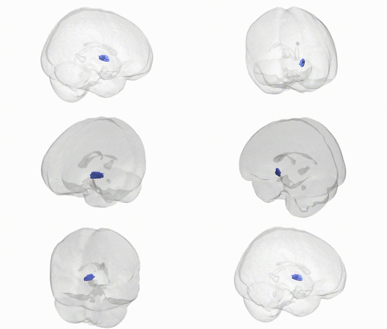
Full Quality Version: Download MP4
Wali Sidiqyar*, Gaurav Rudravaram*, Elyssa M. McMaster, Trent M. Schwartz, Adam M. Saunders, Kurt G. Schilling, Bennett A. Landman "Introducing SPINS: A Shared Public Visualization Library of Neuroanatomical Structures." Medical Imaging with Deep Learning- short paper
This resource is licensed under CC0 1.0 Universal (Public Domain).