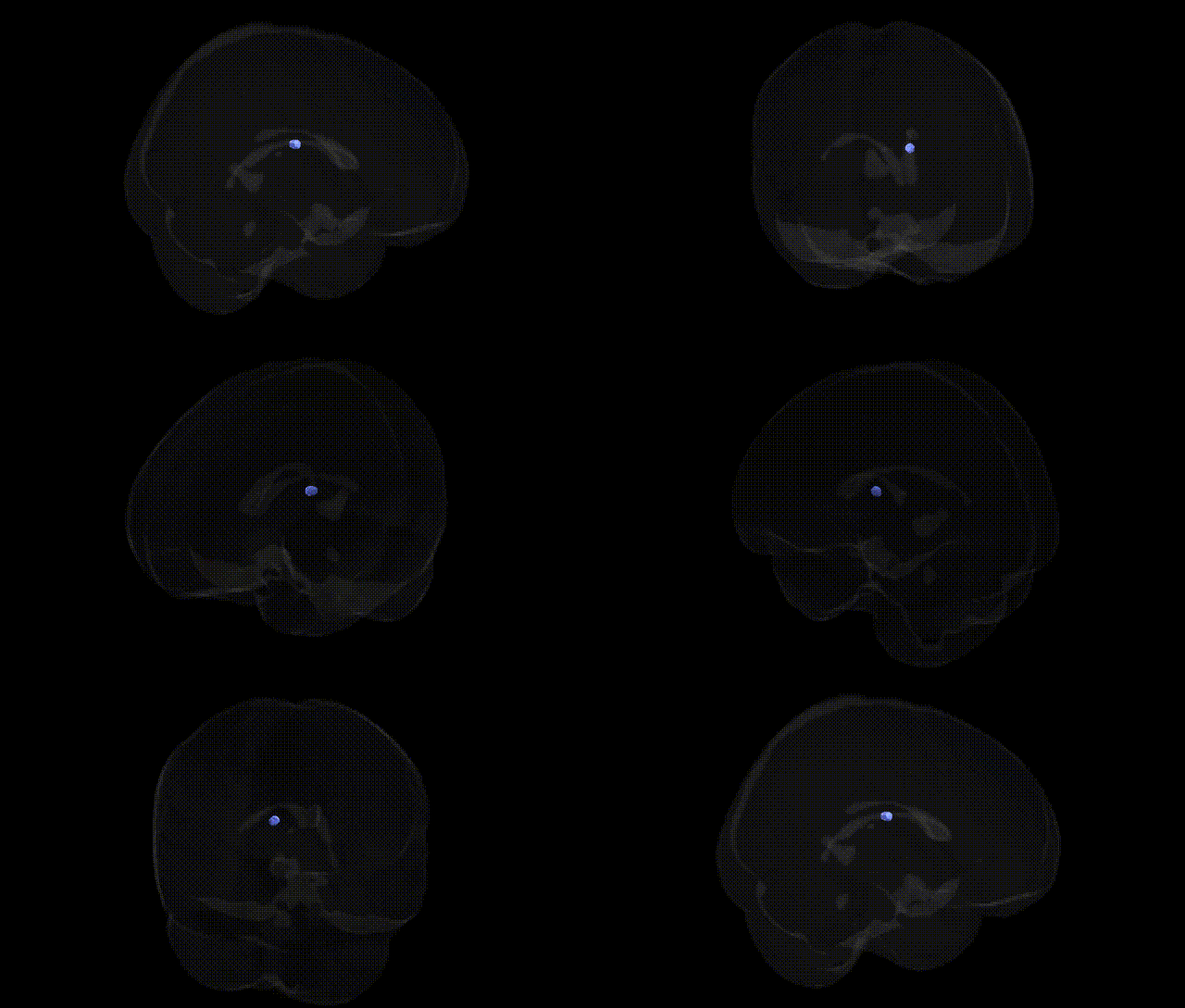
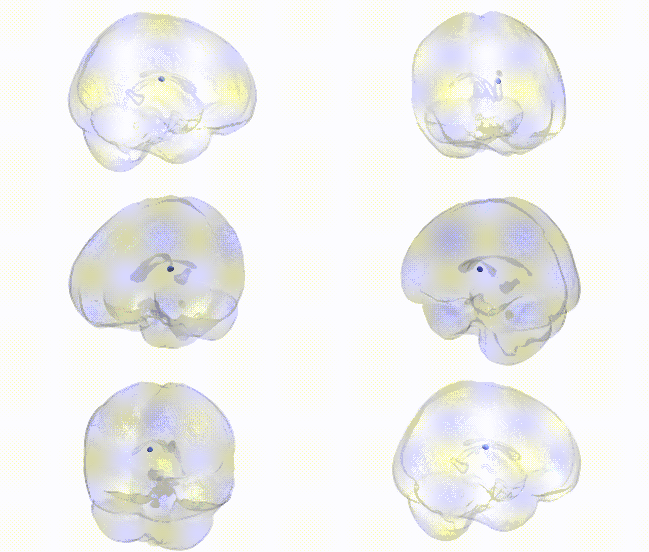
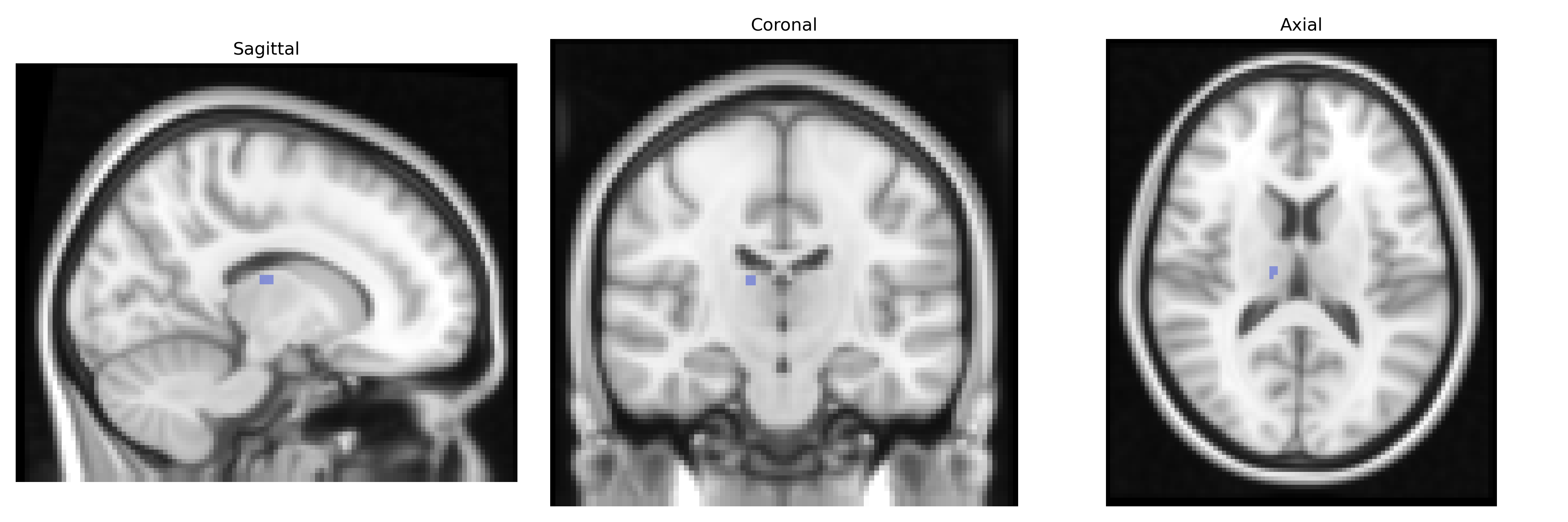
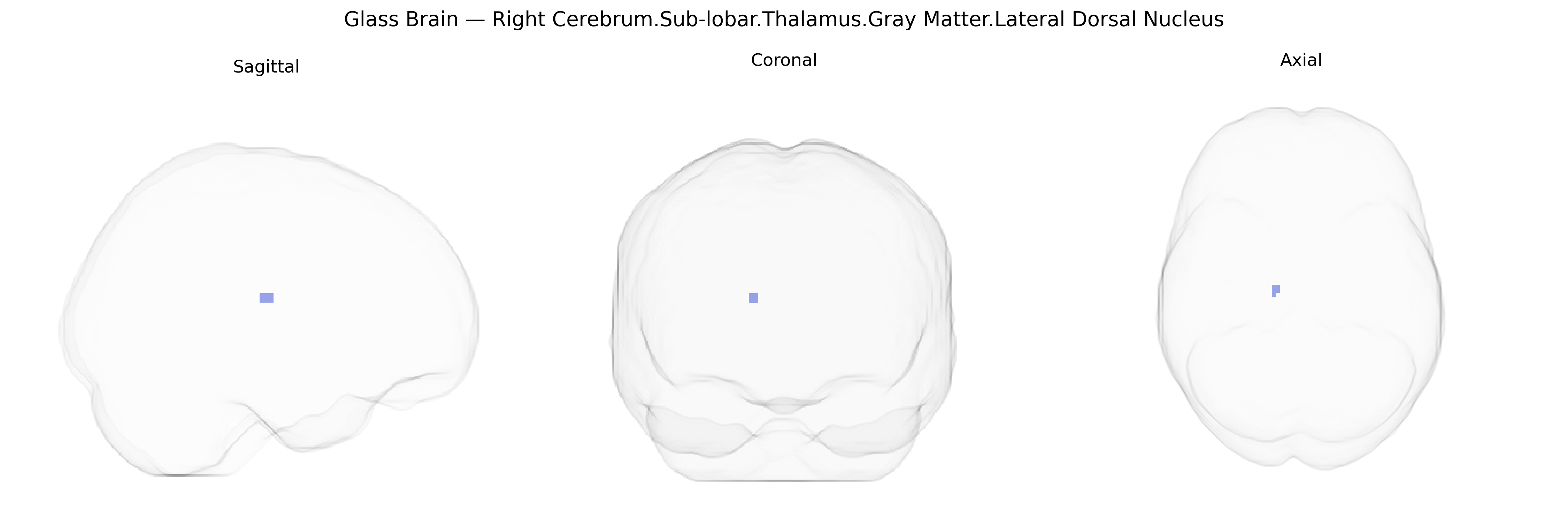
The bilateral Right Cerebrum.Sub-lobar.Thalamus.Gray Matter.Lateral Dorsal Nucleus corresponds to the lateral dorsal (LD) nucleus of the thalamus, a gray matter structure situated in the pulvinar-dorsal tier of thalamic nuclei. The LD nucleus receives multimodal afferents from subcortical structures and cortical regions, including visual and limbic-related areas, and projects primarily to the posterior cingulate and parietal cortices, contributing to visuospatial processing, attention, and aspects of memory and emotional integration. Functionally, it participates in thalamo-cortical circuits that support orientation to environmental stimuli and the integration of sensory information with internal states, and it is often grouped with other association nuclei in functional and structural neuroimaging atlases such as the Talairach 2 mm atlas. There is no direct Wikipedia article for the lateral dorsal nucleus; a closely related structure is the Thalamus.
The lateral dorsal nucleus of the thalamus, a sub-lobar gray-matter structure within the right cerebrum as defined in the Talairach 2 mm atlas, has been most strongly implicated indirectly through genetic associations with thalamic volume and thalamo-cortical network function rather than nucleus-specific GWAS hits. Large-scale imaging–genetics studies (e.g., ENIGMA and UK Biobank) have identified common variants in genes involved in neurodevelopment, synaptic signaling, and axonal guidance—such as those in or near CACNA1C, GRIN2A, BDNF, and MIR137—as associated with global or regional thalamic volume and connectivity, traits that encompass lateral dorsal nucleus anatomy and function. Polygenic risk scores for schizophrenia, bipolar disorder, and major depressive disorder show robust associations with thalamic structure and thalamo-cortical circuitry, and several psychiatric GWAS loci (e.g., in CACNA1C, ZNF804A, and DRD2) have been linked to functional and structural alterations in thalamic relay and association nuclei, including the lateral dorsal complex, in neuroimaging studies. Additional GWAS work on cognitive traits, such as general intelligence, attention, and memory, has implicated variants (e.g., in genes related to glutamatergic signaling and neuroplasticity) that modulate thalamic connectivity with the hippocampus and parietal cortex, circuits for which the lateral dorsal nucleus is a key hub. However, current genetic findings generally reference the thalamus as a whole or large subregional parcels, and there are no widely replicated GWAS results that uniquely and specifically isolate the bilateral lateral dorsal nucleus as the sole anatomical substrate of identified risk variants or trait associations.
Overview generated by GPT-4o (2026).
Region ID: 788
Hemisphere: bilateral
Atlas: Talairach labels 2mm
Full Quality Version: Download MP4
Full Quality Version: Download MP4
Wali Sidiqyar*, Gaurav Rudravaram*, Elyssa M. McMaster, Trent M. Schwartz, Adam M. Saunders, Kurt G. Schilling, Bennett A. Landman "Introducing SPINS: A Shared Public Visualization Library of Neuroanatomical Structures." Medical Imaging with Deep Learning- short paper
This resource is licensed under CC0 1.0 Universal (Public Domain).