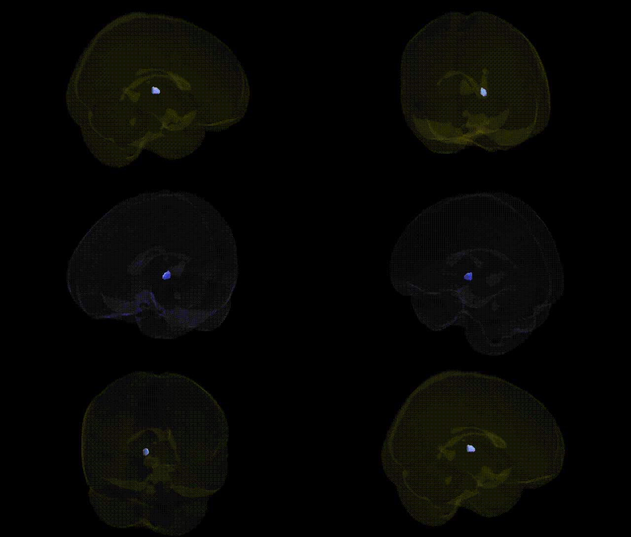
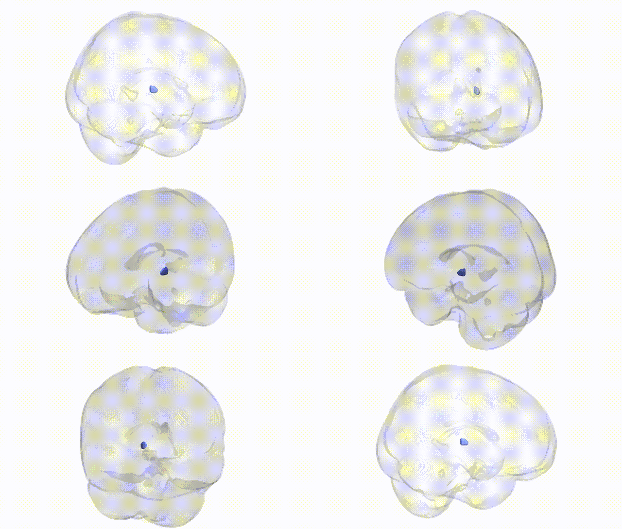
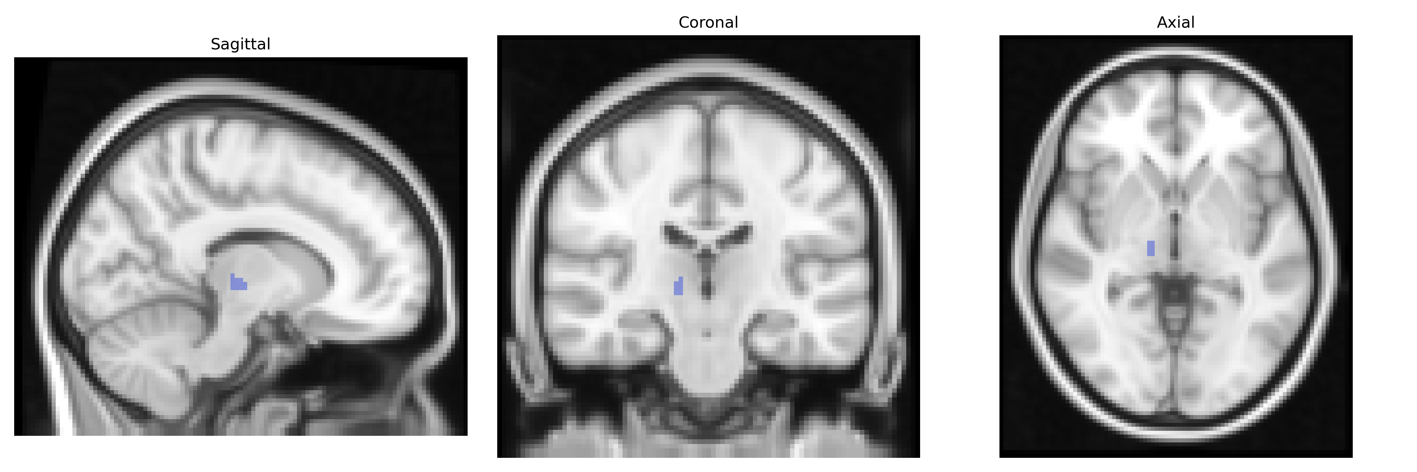
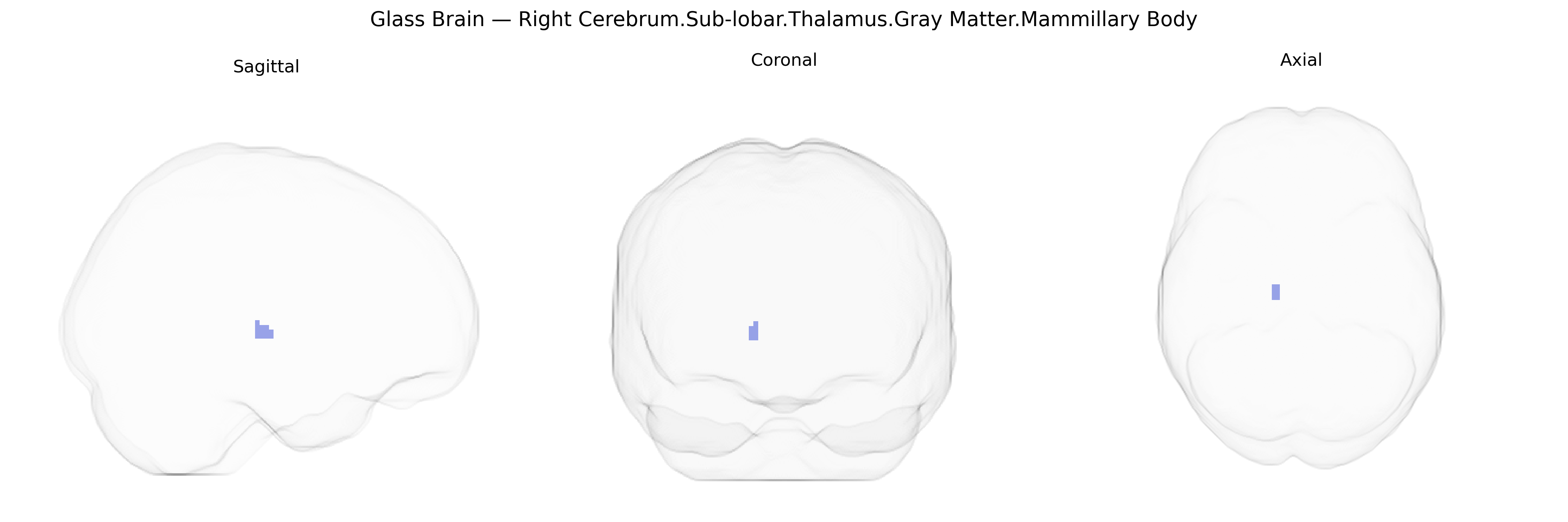
The bilateral Right Cerebrum.Sub-lobar.Thalamus.Gray Matter.Mammillary Body corresponds to a pair of small, rounded nuclei located on the ventral surface of the diencephalon, forming part of the posterior hypothalamus and the limbic system. These mammillary bodies receive major input from the hippocampal formation via the fornix and project primarily to the anterior thalamic nuclei through the mammillothalamic tract, forming a key relay within the Papez circuit involved in episodic memory and spatial navigation. Damage to the mammillary bodies, frequently associated with thiamine deficiency in conditions such as Wernicke–Korsakoff syndrome, is linked to profound anterograde amnesia and confabulation, underscoring their role in memory consolidation. Mammillary body
The mammillary bodies, small hypothalamic structures adjacent to the thalamus, are most strongly implicated in genetic and neurodevelopmental mechanisms underlying memory circuits rather than in large-scale GWAS of region-specific gray matter volume, as most imaging-genetics studies do not isolate this structure at 2 mm Talairach resolution. Genetic associations relevant to the mammillary body region arise primarily from disorders with diencephalic and limbic involvement: variants in thiamine transport and metabolism genes (such as SLC19A2 and others affecting nutritional deficiency risk) contribute indirectly to vulnerability for Wernicke–Korsakoff syndrome, where mammillary body damage is characteristic. Broader GWAS of subcortical and thalamic volumes (e.g., ENIGMA and UK Biobank studies) have identified loci near genes such as DLG2, SLC39A8, and SPTBN1 that influence thalamic and limbic structures, although specific mammillary body volumetric GWAS are lacking. Mendelian conditions and copy-number variants that disrupt hypothalamic–limbic development (including some 22q11.2 deletions and genes like PROKR2 or FGFR1 implicated in hypothalamic anomalies) can alter mammillary body integrity as part of more global diencephalic malformations. In psychiatric and cognitive GWAS, risk loci for schizophrenia, bipolar disorder, and major depression affecting synaptic and plasticity-related genes (e.g., CACNA1C, GRIN2A, and others) are enriched in networks encompassing the hippocampal–mammillary–thalamic memory circuit, linking genetic variation to functional dysconnectivity rather than to this single anatomical label. Overall, current genetic evidence ties the mammillary body region to inherited susceptibility for memory impairment, thiamine-related neurotoxicity, and limbic–thalamic circuit dysfunction, but direct GWAS signals specific to bilateral mammillary body gray matter volume remain sparse.
Overview generated by GPT-4o (2026).
Region ID: 582
Hemisphere: bilateral
Atlas: Talairach labels 2mm
Full Quality Version: Download MP4
Full Quality Version: Download MP4
Wali Sidiqyar*, Gaurav Rudravaram*, Elyssa M. McMaster, Trent M. Schwartz, Adam M. Saunders, Kurt G. Schilling, Bennett A. Landman "Introducing SPINS: A Shared Public Visualization Library of Neuroanatomical Structures." Medical Imaging with Deep Learning- short paper
This resource is licensed under CC0 1.0 Universal (Public Domain).