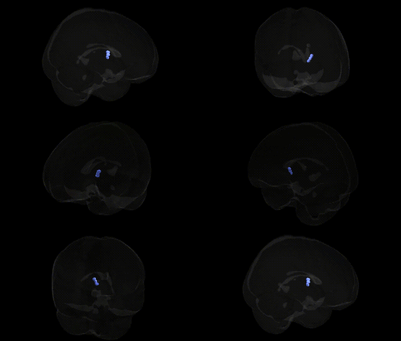
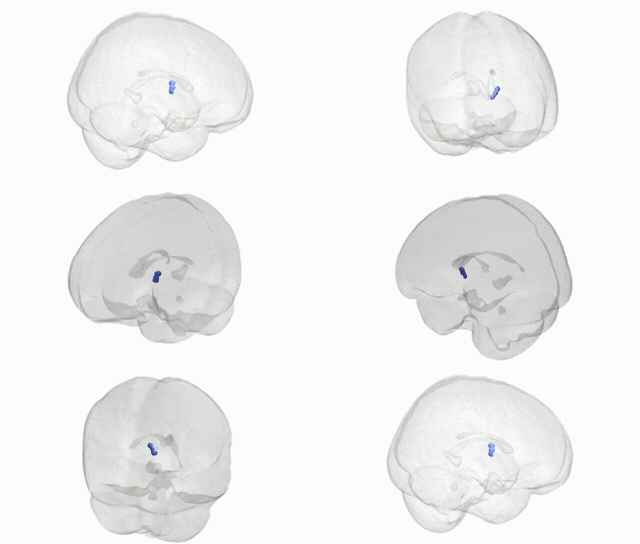
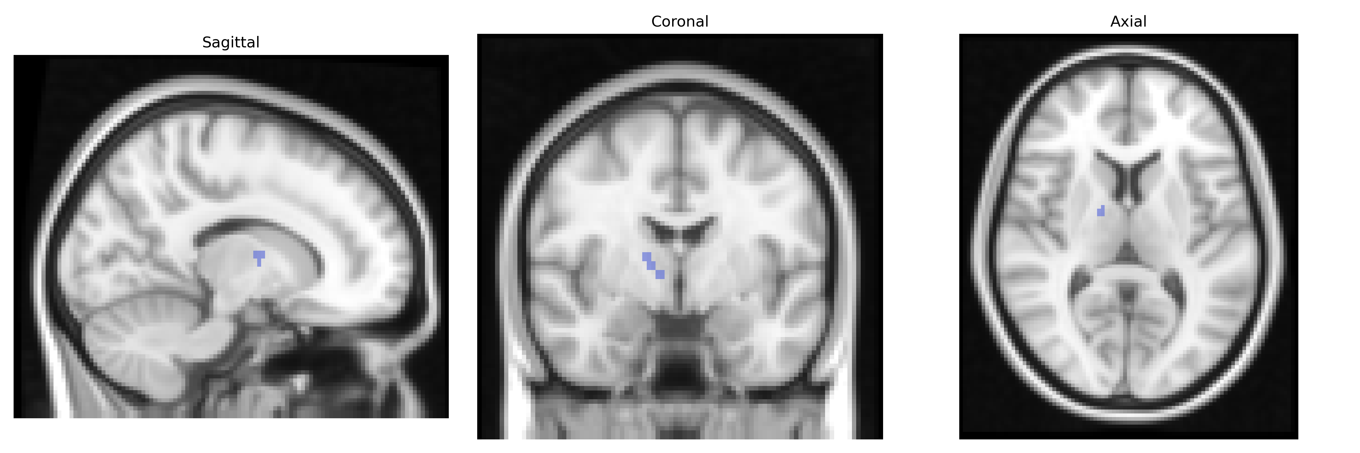
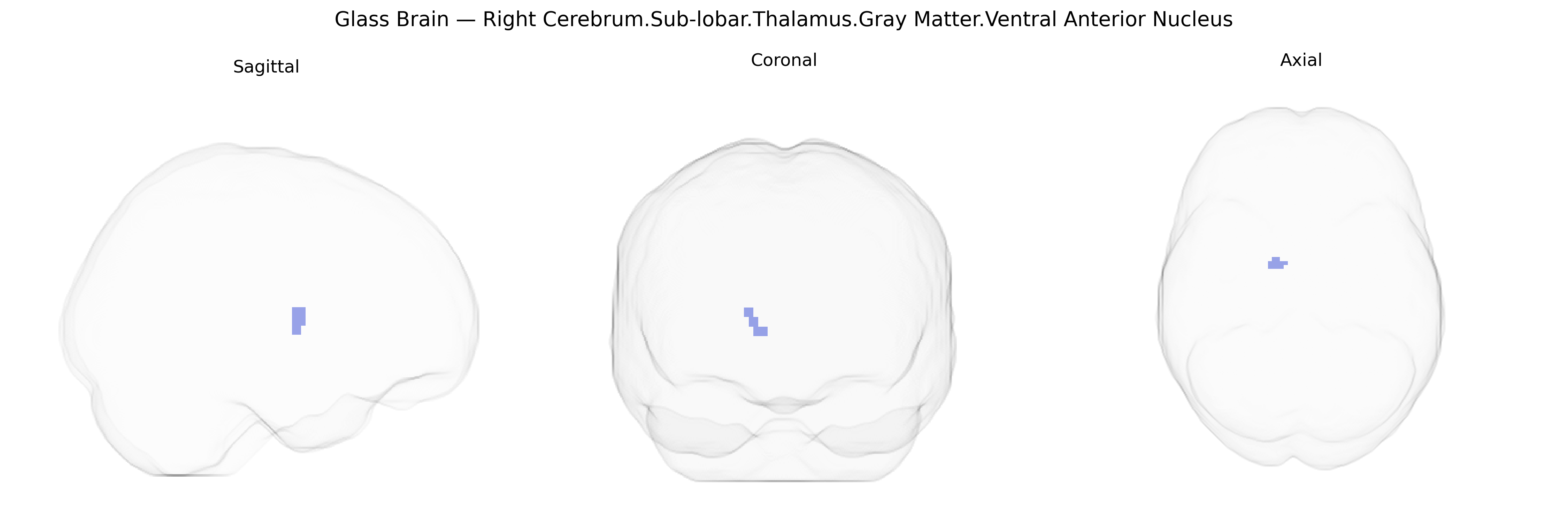
The bilateral Right Cerebrum.Sub-lobar.Thalamus.Gray Matter.Ventral Anterior Nucleus is a thalamic relay nucleus situated in the anterior portion of the ventral thalamus, composed primarily of gray matter and forming part of the sub-lobar structures of the right cerebral hemisphere. Functionally, it receives input mainly from the basal ganglia—especially the globus pallidus and substantia nigra—and projects to premotor and supplementary motor cortical areas, thereby contributing to motor planning, initiation, and the modulation of voluntary movements. It also participates in cortico–basal ganglia–thalamocortical loops implicated in the refinement of motor programs and may have roles in higher-order aspects of action selection and behavioral control. There is no direct link; see the related structure: Thalamus.
The bilateral right ventral anterior nucleus of the thalamus, a sub-lobar gray matter region implicated in motor, cognitive, and limbic integration, has been linked indirectly to several genetic findings through imaging-genetics and GWAS studies of thalamic volume, connectivity, and function, although locus-specific data for this exact Talairach-defined subnucleus are limited. Large GWAS of thalamic volume and thalamic nuclei (e.g., ENIGMA and UK Biobank cohorts) have identified common variants in genes involved in neurodevelopment and synaptic signaling—such as variants near SLC39A8, NPTN, and genes in glutamatergic and GABAergic pathways—that modulate thalamic structure and may encompass the ventral anterior nucleus. Polygenic risk for schizophrenia, bipolar disorder, major depressive disorder, ADHD, and autism spectrum disorder has been associated with thalamic volume and thalamo-cortical connectivity, implicating genetic architectures that influence this region’s morphology and function. Imaging-genetics analyses have further linked thalamic measures, including volumes and resting-state connectivity patterns, to variants in genes such as CACNA1C, GRIN2A, and those regulating calcium channels and synaptic plasticity, which have established GWAS associations with psychiatric disorders and cognitive traits. While no large-scale GWAS has focused exclusively on the ventral anterior nucleus under Talairach 2 mm parcellation, convergent evidence from thalamic-wide genetic studies suggests that the genetic influences affecting thalamic development, sensorimotor integration, and higher-order cognition likely extend to this nucleus, contributing to susceptibility for neuropsychiatric disorders, motor symptoms in conditions such as Parkinson’s disease (e.g., via circuit-level DBS targeting), and interindividual differences in executive function and habit learning.
Overview generated by GPT-4o (2026).
Region ID: 647
Hemisphere: bilateral
Atlas: Talairach labels 2mm
Full Quality Version: Download MP4
Full Quality Version: Download MP4
Wali Sidiqyar*, Gaurav Rudravaram*, Elyssa M. McMaster, Trent M. Schwartz, Adam M. Saunders, Kurt G. Schilling, Bennett A. Landman "Introducing SPINS: A Shared Public Visualization Library of Neuroanatomical Structures." Medical Imaging with Deep Learning- short paper
This resource is licensed under CC0 1.0 Universal (Public Domain).