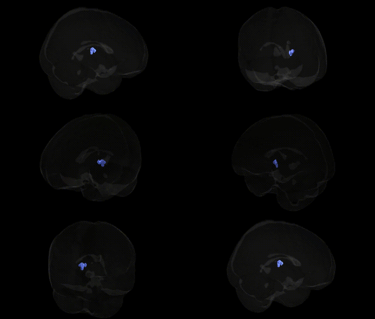
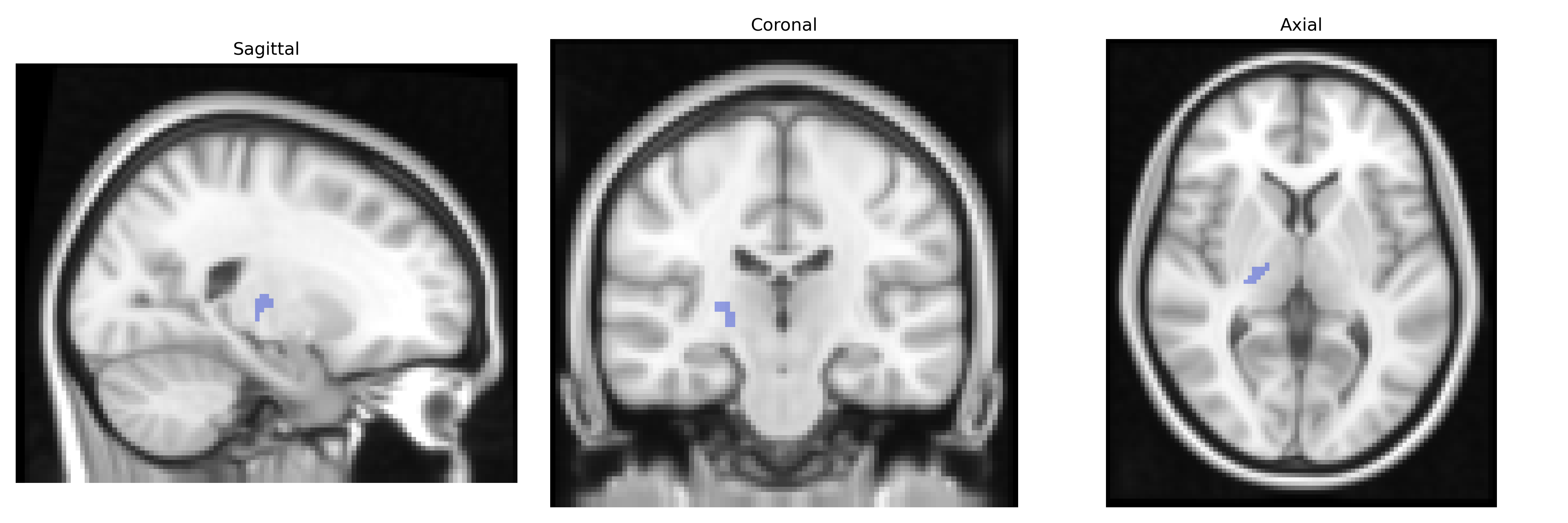
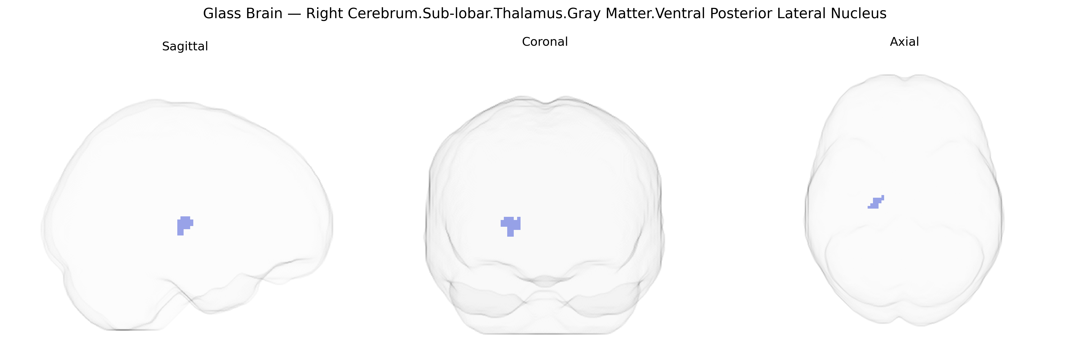
The bilateral Right Cerebrum.Sub-lobar.Thalamus.Gray Matter.Ventral Posterior Lateral Nucleus corresponds to the ventral posterolateral (VPL) nucleus of the thalamus, a key somatosensory relay structure located in the subcortical gray matter of the diencephalon. The VPL receives primary afferent input from the medial lemniscus and spinothalamic tracts, carrying information about touch, vibration, proprioception, pain, and temperature from the body (excluding the face), and projects mainly to the primary somatosensory cortex in the postcentral gyrus. As part of the lateral thalamic nuclei, it plays a critical role in conscious perception of bodily sensation, sensorimotor integration, and modulation of nociceptive signals. Lesions in this region can lead to sensory deficits such as loss or distortion of tactile and pain perception, paresthesias, or central post-stroke pain syndromes. There is no direct Wikipedia article for this specific nucleus; a related structure entry is: Thalamus.
The ventral posterior lateral (VPL) nucleus of the thalamus, a key somatosensory relay region, has been implicated indirectly in genetic studies through its structural and functional properties rather than via locus-specific GWAS hits, as most large-scale analyses focus on broader thalamic volumes or somatosensory networks. Variants in genes related to thalamocortical development and synaptic transmission—such as those affecting glutamatergic and GABAergic signaling (e.g., CACNA1C, GRM3, and other neurodevelopmental genes identified in psychiatric GWAS)—have been associated with altered thalamic structure and connectivity, which includes dorsal and ventral posterior nuclei, in disorders like schizophrenia, bipolar disorder, and major depressive disorder. Neuroimaging genetics consortia (e.g., ENIGMA) have reported heritable influences on thalamic volume and microstructure, with polygenic risk scores for schizophrenia, autism spectrum disorder, and ADHD showing associations with thalamic alterations that encompass somatosensory relay regions. In chronic pain and migraine, GWAS-implicated loci in ion channel and neuroinflammatory genes (such as SCN9A and genes in the CGRP pathway) have been linked to functional and structural changes in thalamic pain-processing circuits, including posterior thalamic nuclei, although not always anatomically resolved to the VPL in Talairach space. Overall, genetic associations point to a broad pattern in which polygenic variation influencing neurodevelopment, synaptic signaling, and pain processing affects thalamic nuclei involved in somatosensory and nociceptive transmission, with the VPL serving as a key node but rarely isolated as a unique GWAS target in current atlases and datasets.
Overview generated by GPT-4o (2026).
Region ID: 584
Hemisphere: bilateral
Atlas: Talairach labels 2mm
Full Quality Version: Download MP4
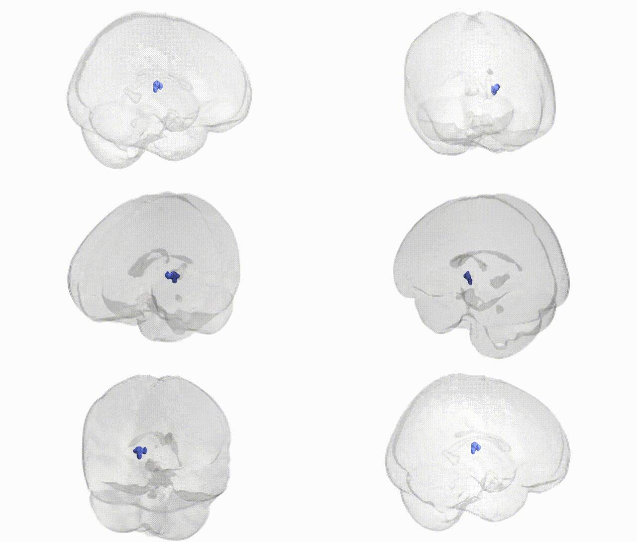
Full Quality Version: Download MP4
Wali Sidiqyar*, Gaurav Rudravaram*, Elyssa M. McMaster, Trent M. Schwartz, Adam M. Saunders, Kurt G. Schilling, Bennett A. Landman "Introducing SPINS: A Shared Public Visualization Library of Neuroanatomical Structures." Medical Imaging with Deep Learning- short paper
This resource is licensed under CC0 1.0 Universal (Public Domain).