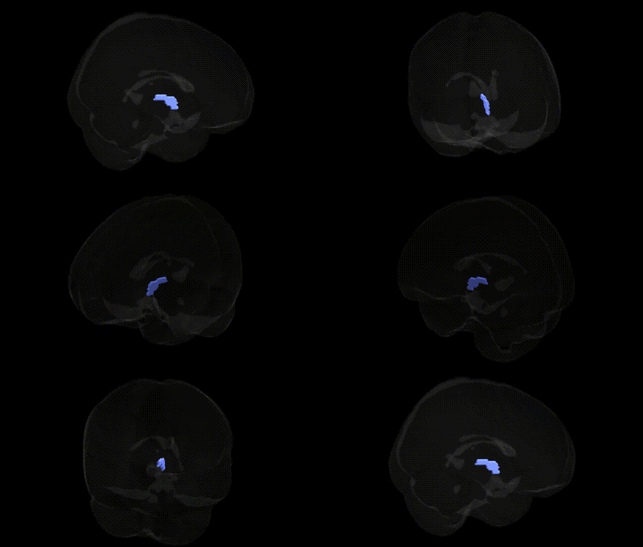
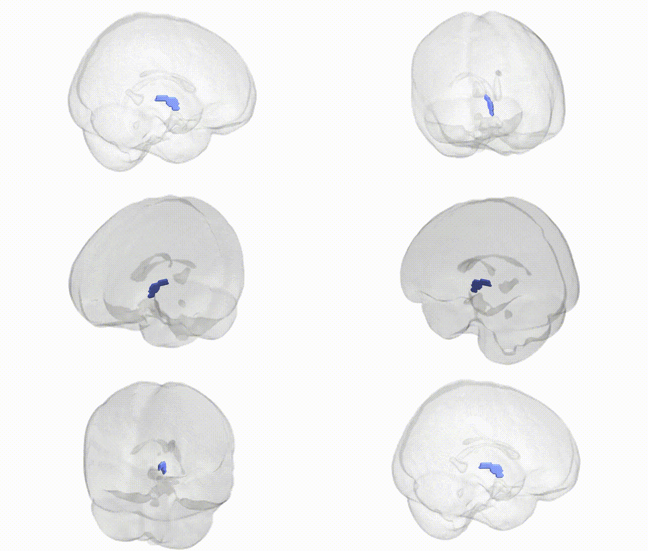
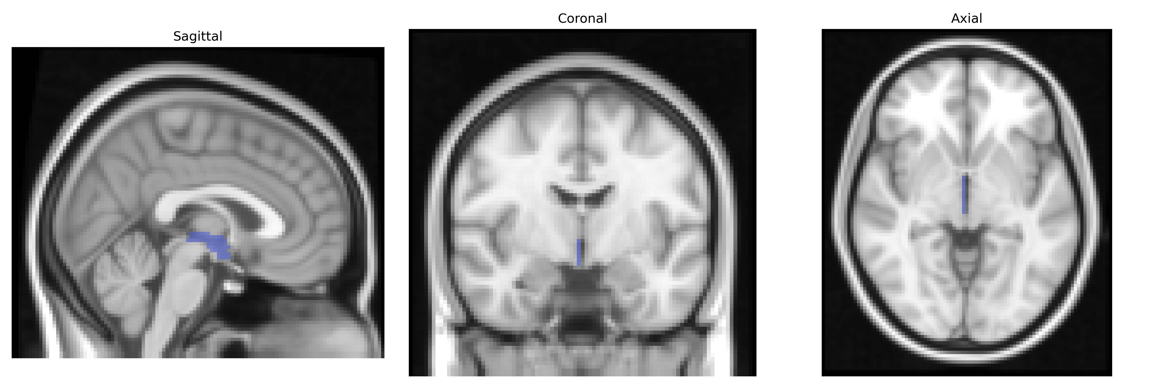
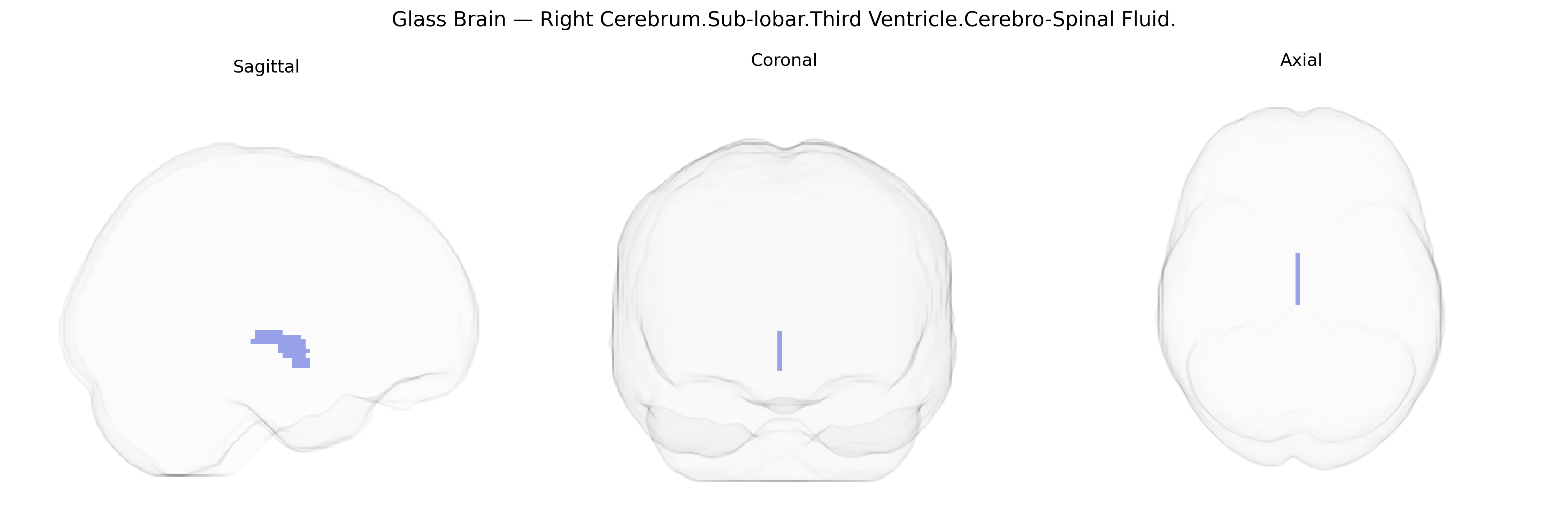
The bilateral Right Cerebrum.Sub-lobar.Third Ventricle.Cerebro-Spinal Fluid region, as defined in the Talairach 2 mm atlas, corresponds to cerebrospinal fluid (CSF) contained within the third ventricle, a narrow midline cavity situated between the thalami and hypothalamus in the diencephalon. This ventricular space is part of the brain’s CSF circulation pathway, receiving fluid from the lateral ventricles via the interventricular foramina and draining into the cerebral aqueduct toward the fourth ventricle. The third ventricle contributes to maintaining intracranial pressure, providing mechanical protection, and supporting metabolic exchange and homeostasis for surrounding deep gray matter structures. Although CSF itself is a fluid rather than neural tissue, its distribution within the third ventricle is crucial for normal brain function and can be altered in conditions such as hydrocephalus, tumors, or inflammatory processes affecting the ventricular system.
Genetic associations specific to the bilateral Right Cerebrum.Sub-lobar.Third Ventricle.Cerebro-Spinal Fluid region, as defined by the Talairach 2 mm atlas, are generally indirect, reflecting its role as a cerebrospinal fluid (CSF)-filled midline structure rather than gray matter. Large neuroimaging GWAS consortia such as ENIGMA and UK Biobank have identified numerous loci associated with total and regional ventricular volume, with variants near or within genes including APOE, MAPT, BIN1, SORL1, and CR1 repeatedly implicated in enlargement of third and lateral ventricles, largely via neurodegenerative and atrophic processes. Ventricular size has been genetically linked to Alzheimer’s disease, Parkinson’s disease, schizophrenia, bipolar disorder, and major depressive disorder, as well as general cognitive performance and brain aging, suggesting shared genetic architecture between ventricular CSF volume and these traits. In particular, APOE ε4 and other Alzheimer’s risk alleles correlate with greater third-ventricle and global ventricular volumes, while schizophrenia and bipolar disorder risk variants show polygenic associations with enlarged ventricles and altered CSF spaces. Additional GWAS have related ventricular and CSF measures to loci involved in neurodevelopment, axonal guidance, and extracellular matrix (e.g., genes near 3p24–p25 and 10q26), reflecting developmental influences on ventricular morphology. Overall, genetic findings link the third ventricle’s CSF volume to polygenic risk for neurodegenerative and psychiatric disorders, brain atrophy, and cognitive decline, rather than to region-specific functional specialization.
Overview generated by GPT-4o (2026).
Region ID: 360
Hemisphere: bilateral
Atlas: Talairach labels 2mm
Full Quality Version: Download MP4
Full Quality Version: Download MP4
Wali Sidiqyar*, Gaurav Rudravaram*, Elyssa M. McMaster, Trent M. Schwartz, Adam M. Saunders, Kurt G. Schilling, Bennett A. Landman "Introducing SPINS: A Shared Public Visualization Library of Neuroanatomical Structures." Medical Imaging with Deep Learning- short paper
This resource is licensed under CC0 1.0 Universal (Public Domain).