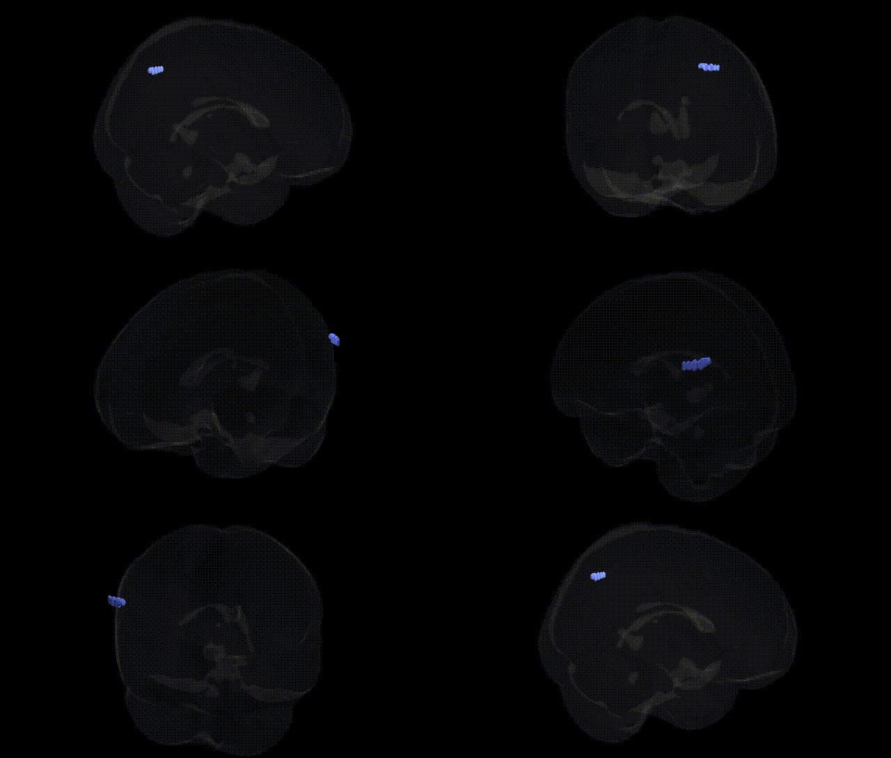
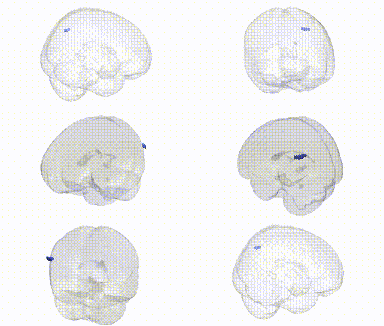
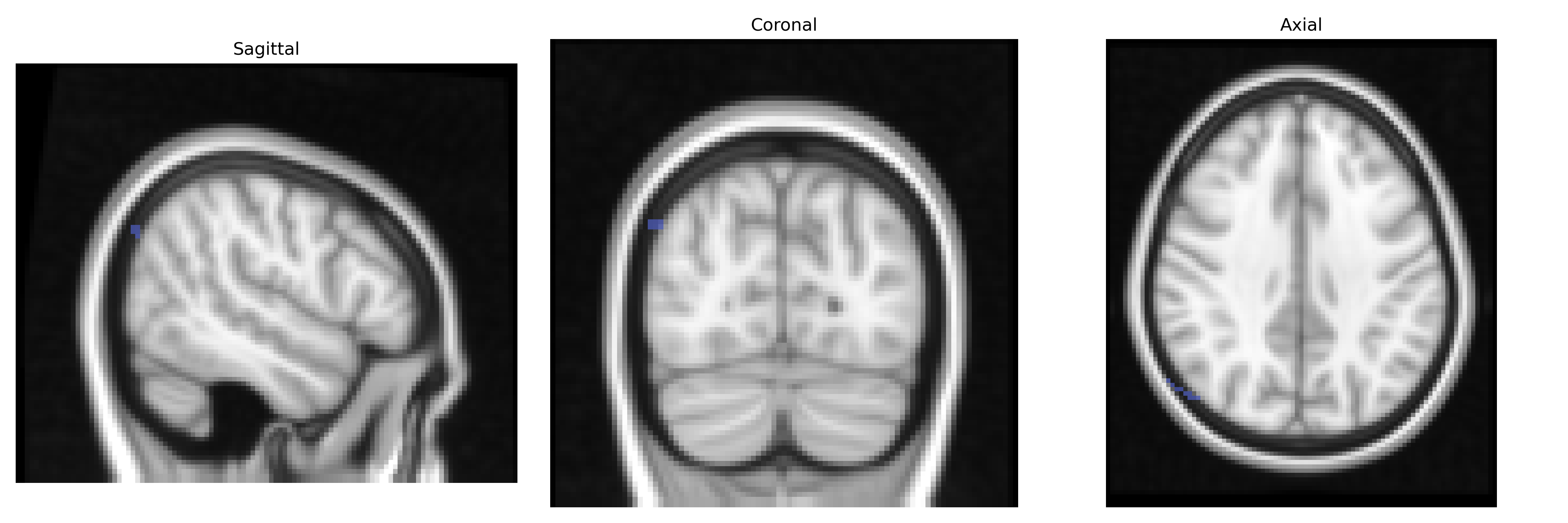
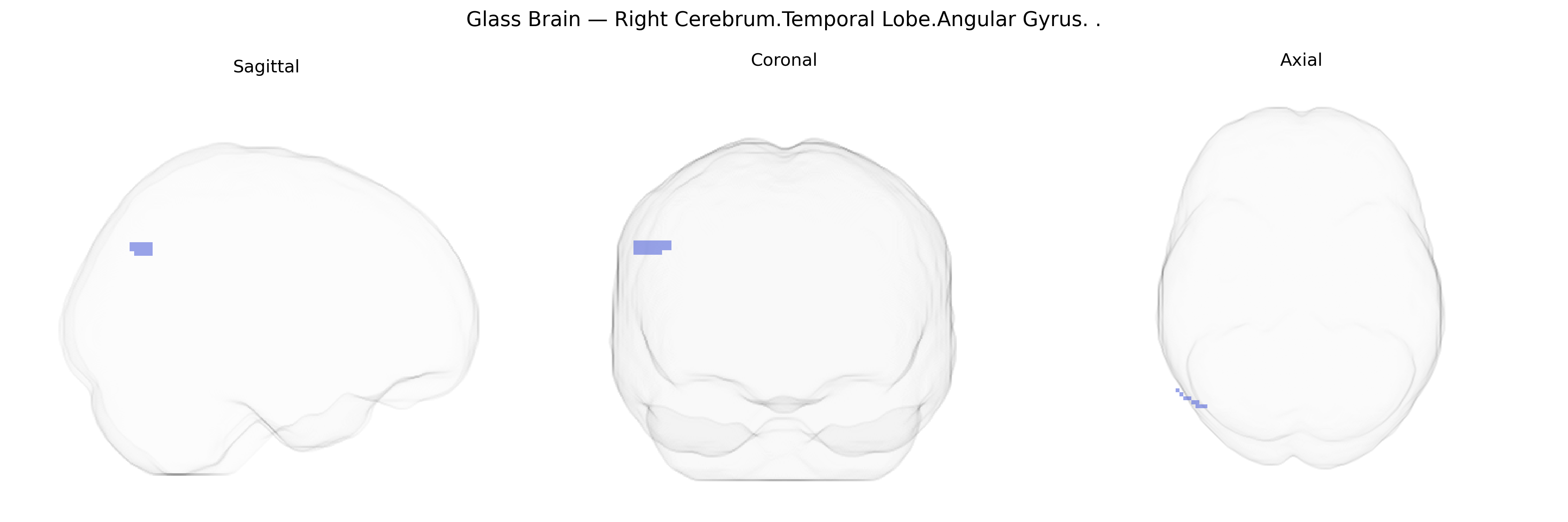
The bilateral right angular gyrus of the temporal lobe is a multimodal association area located at the junction of the temporal, parietal, and occipital lobes, corresponding roughly to Brodmann area 39. It integrates visual, auditory, and somatosensory information and plays a key role in higher cognitive functions including language comprehension, reading, number processing, spatial cognition, memory retrieval, and aspects of theory of mind. This region is heavily interconnected with other association cortices and limbic structures, supporting semantic processing and the integration of conceptual knowledge; lesions here are associated with Gerstmann syndrome, alexia, agraphia, and disturbances in spatial and attentional processing. Angular gyrus
The angular gyrus, a heteromodal association area in the temporal–parietal junction, has been implicated in multiple genetically influenced traits and disorders, although GWAS rarely target the precise Talairach-defined bilateral Right Cerebrum.Temporal Lobe.Angular Gyrus label. Large neuroimaging GWAS (e.g., ENIGMA and UK Biobank) have identified common variants near genes such as HMGA2, MIR137, KIAA0586, and others that influence cortical thickness, surface area, and gyrification in inferior parietal and temporoparietal regions overlapping the angular gyrus, suggesting polygenic control of its structural variation. Functionally, this region has been repeatedly associated with reading and language, number processing, semantic integration, and default mode network activity, and genetic risk for dyslexia, developmental language disorder, and mathematical ability overlaps with pathways affecting temporoparietal cortex (e.g., ROBO1, DCDC2, KIAA0319, FOXP2-related networks), although these are not angular-gyrus–specific. Psychiatric and neurodevelopmental GWAS for schizophrenia, autism spectrum disorder, ADHD, and major depression show enrichment of risk variants in genes involved in synaptic plasticity and neurodevelopment (such as CACNA1C, GRIN2A, CNTNAP2), and imaging–genetics studies often report altered activation or connectivity of the right angular gyrus within default mode and attention networks in carriers of high polygenic risk. Additionally, APOE and other Alzheimer’s disease risk variants are associated with atrophy and hypometabolism in temporoparietal regions including the angular gyrus, which serves as a key hub affected in early Alzheimer’s and other dementias. Overall, genetic influences on this region are highly polygenic and shared across large-scale networks, with no single gene uniquely defining the bilateral right angular gyrus but multiple loci contributing to its morphology, connectivity, and vulnerability to cognitive and psychiatric phenotypes.
Overview generated by GPT-4o (2026).
Region ID: 960
Hemisphere: bilateral
Atlas: Talairach labels 2mm
Full Quality Version: Download MP4
Full Quality Version: Download MP4
Wali Sidiqyar*, Gaurav Rudravaram*, Elyssa M. McMaster, Trent M. Schwartz, Adam M. Saunders, Kurt G. Schilling, Bennett A. Landman "Introducing SPINS: A Shared Public Visualization Library of Neuroanatomical Structures." Medical Imaging with Deep Learning- short paper
This resource is licensed under CC0 1.0 Universal (Public Domain).