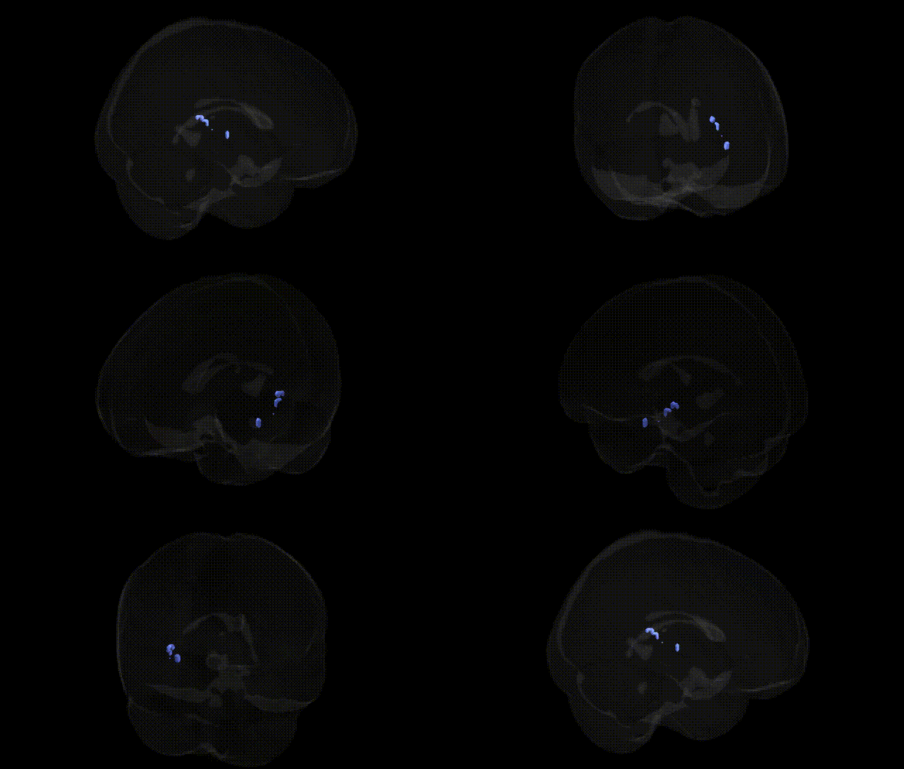
The bilateral Right Cerebrum.Temporal Lobe.Caudate.Gray Matter.Caudate Tail refers to the posterior segment of the caudate nucleus situated within or adjacent to the temporal lobe, composed primarily of gray matter containing medium spiny GABAergic neurons. As part of the dorsal striatum, the caudate tail receives input from association and limbic cortices and contributes to cortico–basal ganglia–thalamocortical loops implicated in sensorimotor integration, procedural learning, habit formation, and aspects of visual and emotional processing. Its position along the temporal horn of the lateral ventricle and proximity to structures such as the hippocampus and amygdala supports roles in memory-guided behavior and affective modulation, while connections with dopaminergic midbrain nuclei integrate reward-related and motivational signals. There is no direct Wikipedia article for the caudate tail; see the related structure Caudate nucleus.
The bilateral right temporal lobe caudate tail gray matter (as localized in the Talairach 2 mm atlas) lies within the broader striatal circuitry, and genetic associations tend to be reported at the level of the caudate nucleus or temporal–striatal networks rather than this subregion specifically. GWAS and imaging genetics studies have repeatedly implicated variants in genes affecting dopaminergic signaling (e.g., DRD2, DARPP-32/PPP1R1B, COMT) and synaptic plasticity (e.g., BDNF, GRIN2B) in caudate volume and function, with downstream relevance to disorders such as schizophrenia, attention-deficit/hyperactivity disorder, obsessive–compulsive disorder, Tourette syndrome, and substance use disorders, where altered caudate structure or connectivity is often observed. Large-scale neuroimaging GWAS (e.g., ENIGMA, UK Biobank) have identified multiple loci associated with striatal (including caudate) gray matter volume—such as variants near PDE1A, DCC, and genes involved in neurodevelopment, axon guidance, and synaptic organization—some of which also associate with cognitive performance, educational attainment, and risk for neuropsychiatric conditions. Temporal–striatal circuits incorporating the caudate tail have further been linked, via polygenic risk scores and gene–brain–behavior analyses, to traits including habit formation, reward learning, and memory processes, with overlapping genetic architectures between caudate structure, general cognitive ability, and psychiatric risk, though highly specific GWAS or candidate-gene findings targeting the caudate tail region defined by Talairach labels remain limited.
Overview generated by GPT-4o (2026).
Region ID: 350
Hemisphere: bilateral
Atlas: Talairach labels 2mm
Full Quality Version: Download MP4
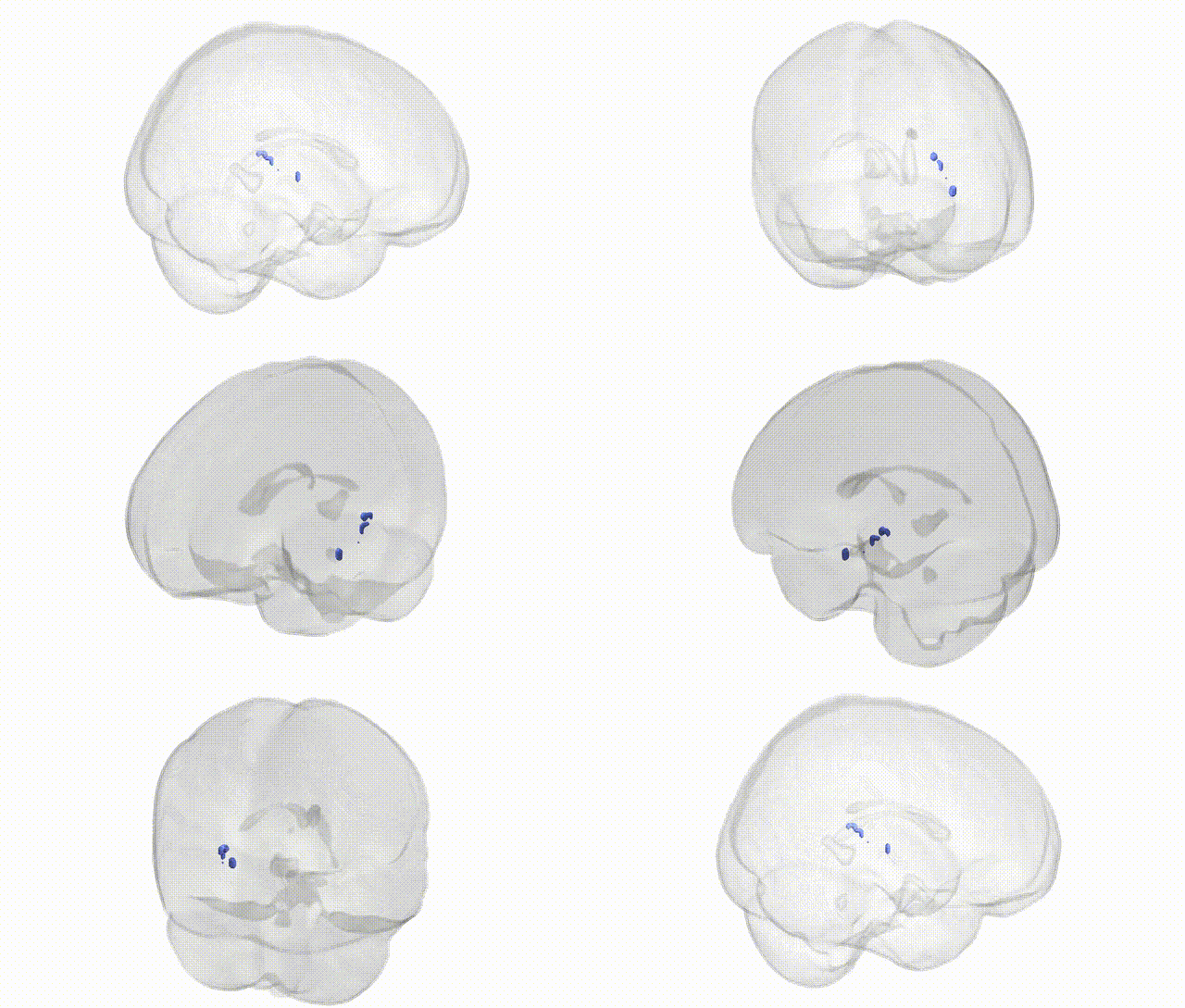
Full Quality Version: Download MP4
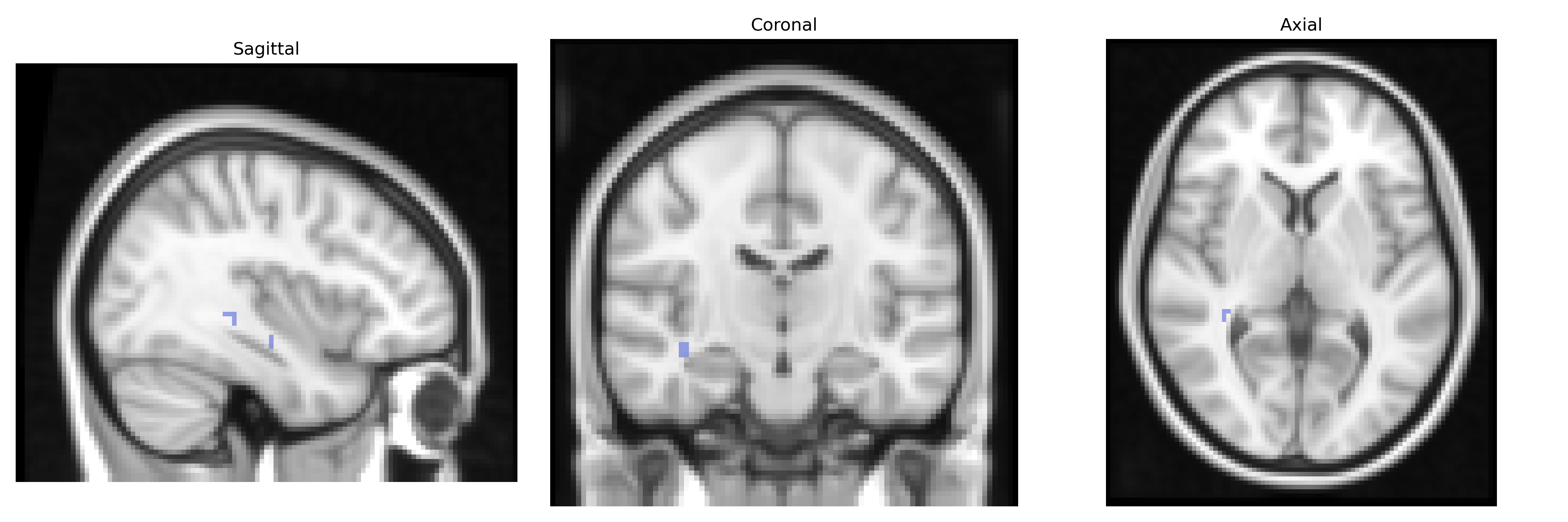
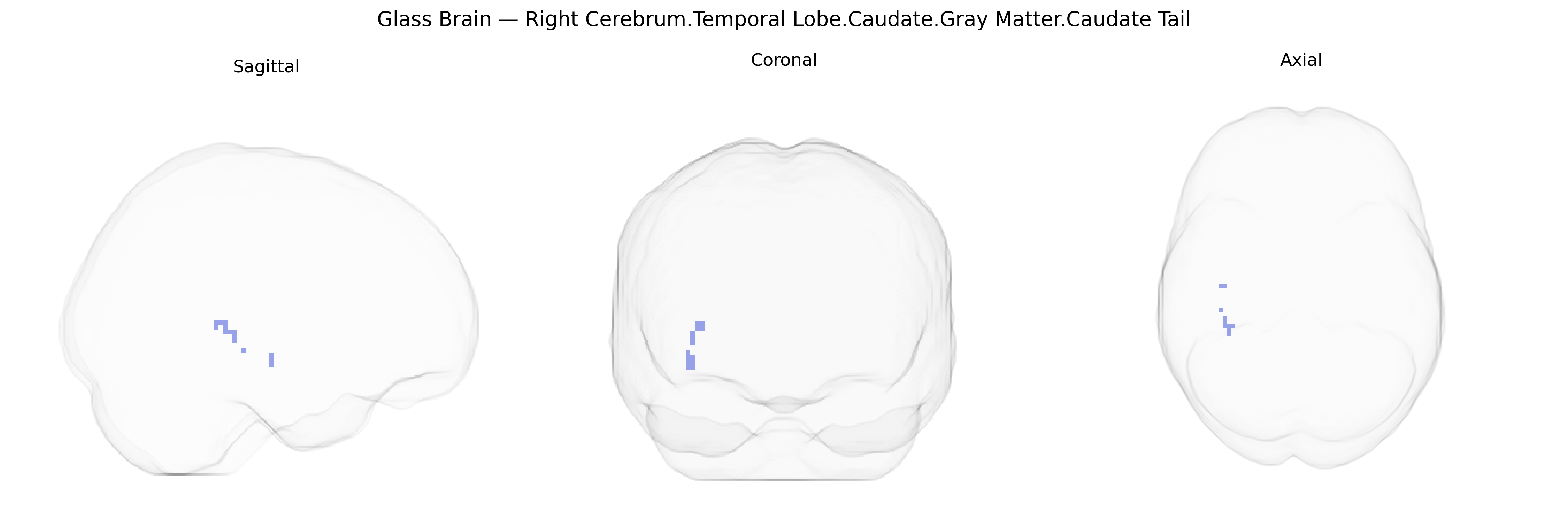
Wali Sidiqyar*, Gaurav Rudravaram*, Elyssa M. McMaster, Trent M. Schwartz, Adam M. Saunders, Kurt G. Schilling, Bennett A. Landman "Introducing SPINS: A Shared Public Visualization Library of Neuroanatomical Structures." Medical Imaging with Deep Learning- short paper
This resource is licensed under CC0 1.0 Universal (Public Domain).