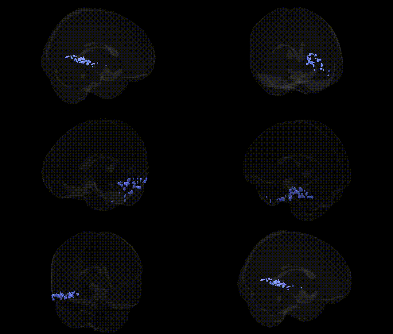
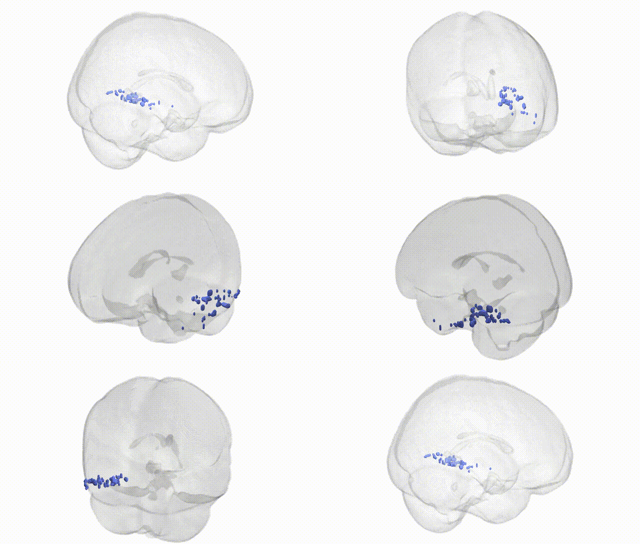
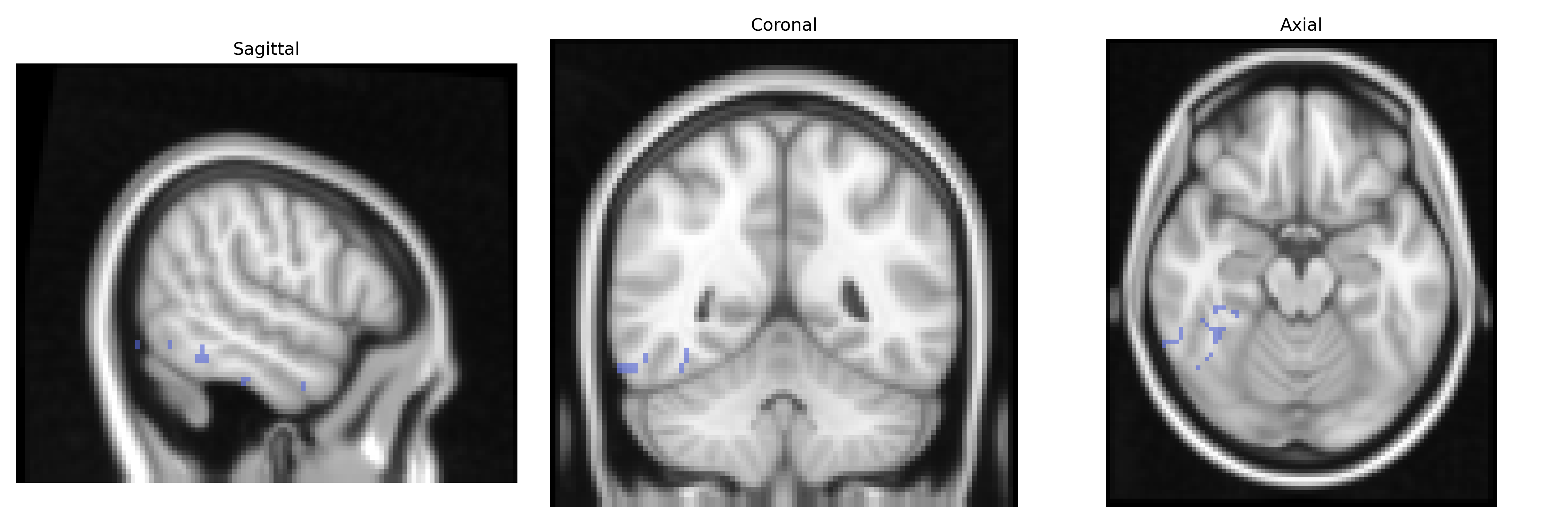
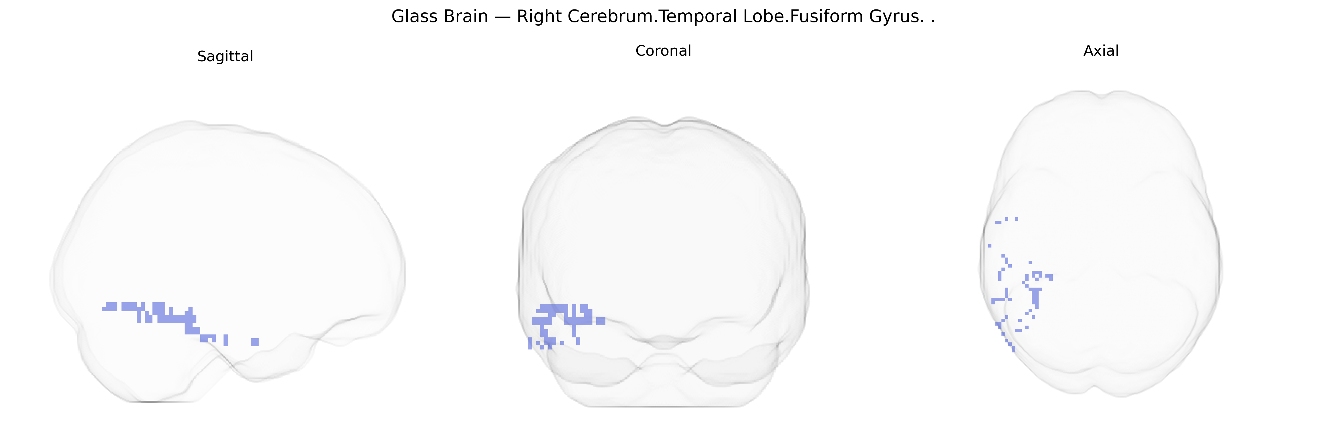
The bilateral Right Cerebrum.Temporal Lobe.Fusiform Gyrus corresponds to cortical tissue on the ventral surface of the temporal lobe, extending from the occipitotemporal junction anteriorly toward the temporal pole, and is part of the ventral visual processing stream. This region is cytoarchitectonically heterogeneous (including areas such as FG1–FG4) and is crucial for high-level visual recognition, including processing of complex stimuli like faces, words, and category-specific objects, as well as contributing to aspects of semantic memory and visual imagery. Its neural activity is strongly modulated by expertise and familiarity with visual categories, and lesions or dysfunction in this area can lead to deficits such as prosopagnosia (impaired face recognition) or acquired dyslexia. Functionally, the fusiform gyrus is extensively connected with occipital visual areas, the parietal cortex, and anterior temporal and prefrontal regions, supporting integrated perception and recognition in multimodal networks.
Fusiform gyrus
The bilateral right fusiform gyrus in the temporal lobe has been implicated in multiple genetic and GWAS findings, largely through imaging genetics studies linking common variants to regional structure and function. Variants in genes related to synaptic development and neuroplasticity (e.g., BDNF, particularly Val66Met) have been associated with fusiform gray matter volume and functional activation in face and object processing tasks, while genome-wide studies of cortical morphology (ENIGMA and related consortia) have identified polygenic influences on fusiform thickness and surface area, including loci near genes involved in neurodevelopmental signaling and cell adhesion. The fusiform gyrus is repeatedly implicated in autism spectrum disorder, where risk variants in genes such as CNTNAP2, SHANK3, and others affect fusiform activation and connectivity during social and face perception, and in prosopagnosia-related studies where familial clustering and linkage suggest heritable influences on fusiform face-selective responses. Schizophrenia GWAS and imaging genetics have linked polygenic risk scores to altered fusiform volume and activation, particularly in social cognition tasks, while genetic risk for dyslexia and language-related traits has been associated with structural and functional differences in ventral temporal regions including fusiform. Large GWAS of cognitive ability and educational attainment show that polygenic scores predict variability in fusiform structure as part of broader temporo-parietal networks, and neurodegenerative disease genetics (e.g., APOE in Alzheimer’s disease) has been related to early atrophy and hypometabolism in fusiform regions tied to visual recognition. Overall, genetic influences on the fusiform gyrus appear highly polygenic, overlapping with risk architectures for neurodevelopmental and psychiatric disorders and reflecting shared mechanisms in cortical development, synaptic function, and visual-social processing.
Overview generated by GPT-4o (2026).
Region ID: 122
Hemisphere: bilateral
Atlas: Talairach labels 2mm
Full Quality Version: Download MP4
Full Quality Version: Download MP4
Wali Sidiqyar*, Gaurav Rudravaram*, Elyssa M. McMaster, Trent M. Schwartz, Adam M. Saunders, Kurt G. Schilling, Bennett A. Landman "Introducing SPINS: A Shared Public Visualization Library of Neuroanatomical Structures." Medical Imaging with Deep Learning- short paper
This resource is licensed under CC0 1.0 Universal (Public Domain).