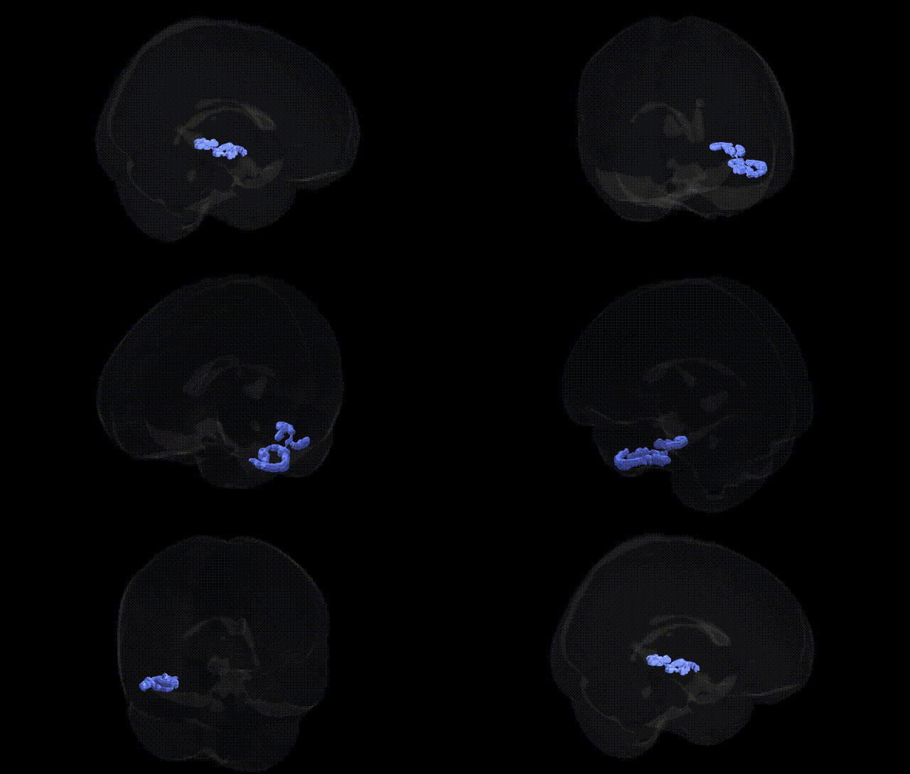
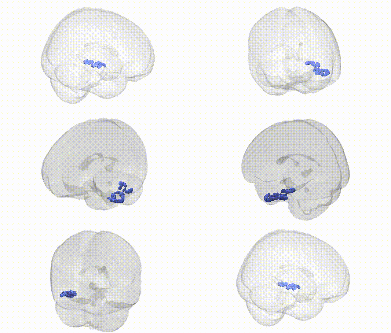
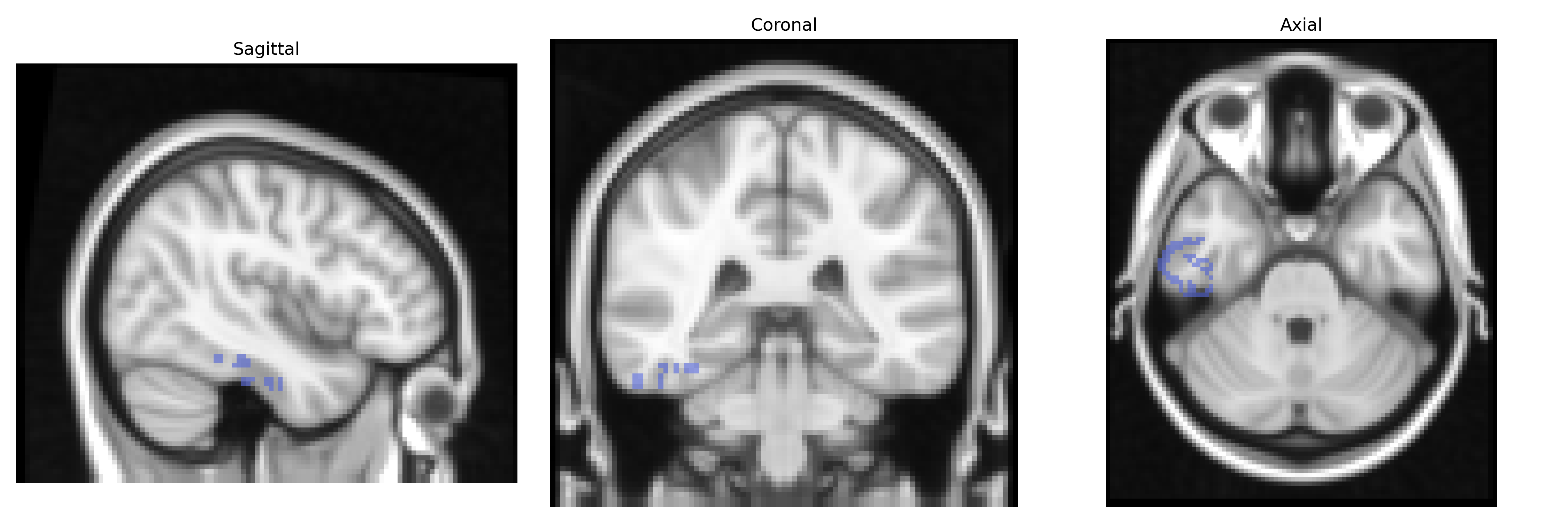
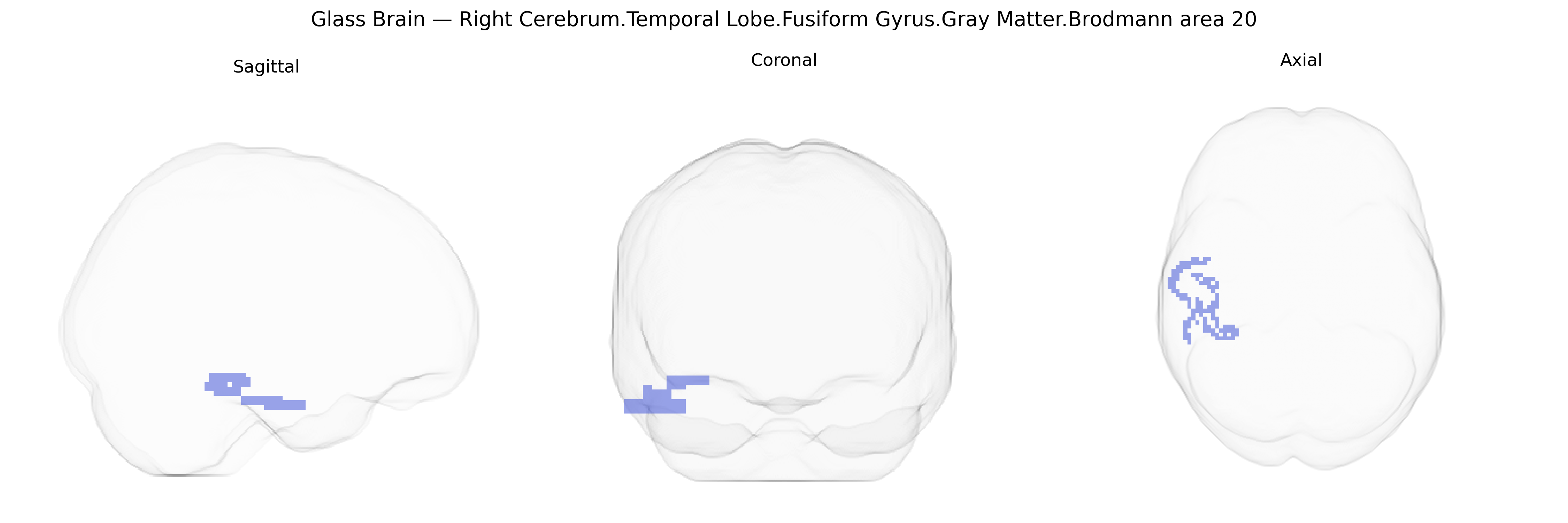
The bilateral Right Cerebrum.Temporal Lobe.Fusiform Gyrus.Gray Matter.Brodmann area 20 corresponds to a cortical region in the inferior temporal portion of the fusiform gyrus, implicated primarily in high-level visual processing and object recognition. As part of the ventral visual stream, BA20 contributes to detailed analysis of complex shapes, faces, and semantic attributes of visual stimuli, integrating visual input with stored memory representations. It is cytoarchitectonically defined by a granular cortex with characteristic laminar organization suited for hierarchical visual and associative processing, and functionally connected to adjacent temporal areas (e.g., BA37, BA21) and limbic regions involved in memory and emotion. Activation in this region is commonly observed during tasks demanding recognition, categorization, and the retrieval of visual object knowledge. There is no direct Wikipedia article for Brodmann area 20 in the fusiform gyrus; a closely related and encompassing structure is the fusiform gyrus: Fusiform gyrus.
The bilateral right fusiform gyrus gray matter in Brodmann area 20, as defined in the Talairach 2mm atlas, has been implicated in several genetic and genome-wide association study (GWAS) findings, particularly in relation to neurodevelopmental and neuropsychiatric traits, as well as brain structural variation. Imaging genetics studies have linked common variants in genes involved in synaptic function and neurodevelopment—such as CNTNAP2, NRXN1, and DISC1—to altered fusiform gyrus volume or activity, especially in the context of autism spectrum disorder, where fusiform structure and activation during face processing are frequently abnormal and heritable. GWAS of brain morphology have identified multiple loci associated with temporal and fusiform cortical thickness and surface area, often mapping to genes regulating neuronal migration, axon guidance, and cortical patterning (for example, variants near PAX6 and other developmental regulators), though these associations are usually reported for broader temporal or ventral visual regions rather than BA20 specifically. Schizophrenia and bipolar disorder risk variants, including those in the MHC region and CACNA1C, have been associated with altered temporal lobe gray matter, with some imaging-genetics analyses reporting fusiform involvement in face recognition and social cognition deficits. Additionally, GWAS of reading ability, dyslexia, and face recognition ability have highlighted genetic contributions to individual differences in ventral occipitotemporal and fusiform function, consistent with the region’s role in high-level visual and language processing. Overall, current genetic evidence supports a polygenic influence on fusiform gyrus structure and function, with BA20 participating in circuits affected in autism, schizophrenia, mood disorders, and cognitive traits, although few studies provide region-specific GWAS signals restricted precisely to bilateral right BA20 as labeled in Talairach.
Overview generated by GPT-4o (2026).
Region ID: 124
Hemisphere: bilateral
Atlas: Talairach labels 2mm
Full Quality Version: Download MP4
Full Quality Version: Download MP4
Wali Sidiqyar*, Gaurav Rudravaram*, Elyssa M. McMaster, Trent M. Schwartz, Adam M. Saunders, Kurt G. Schilling, Bennett A. Landman "Introducing SPINS: A Shared Public Visualization Library of Neuroanatomical Structures." Medical Imaging with Deep Learning- short paper
This resource is licensed under CC0 1.0 Universal (Public Domain).