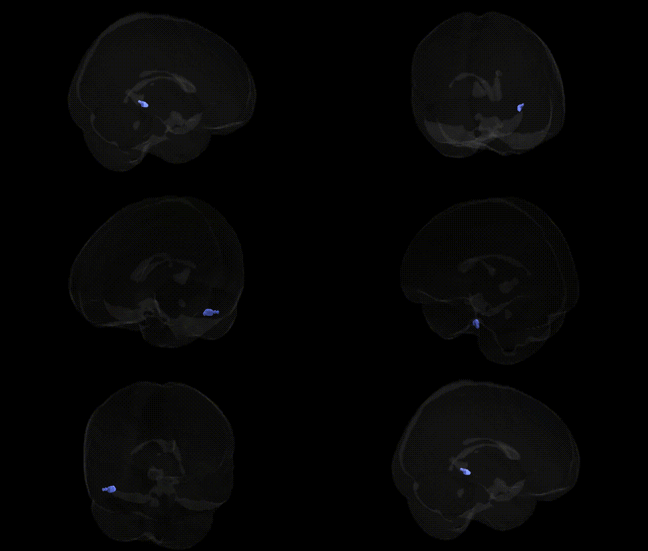
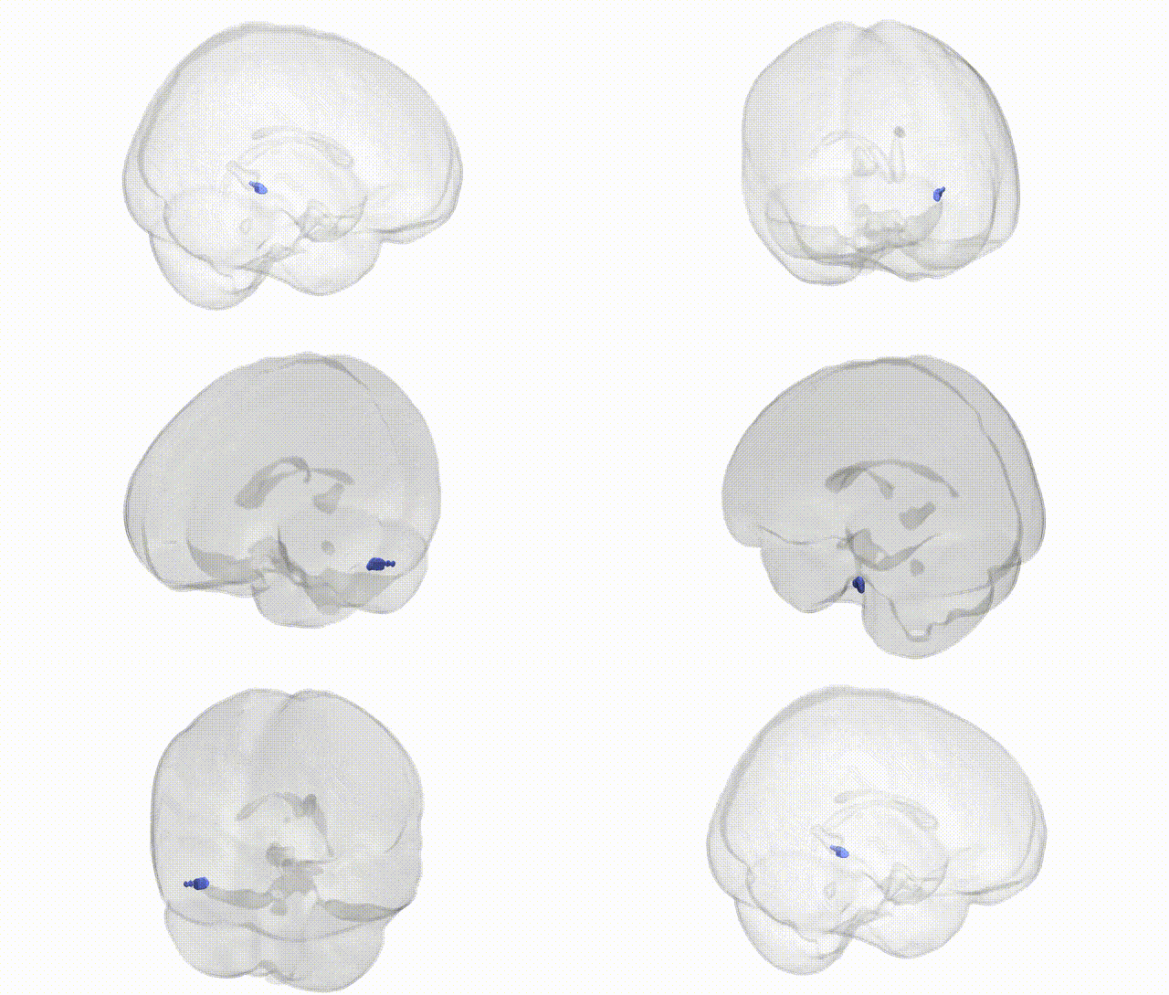
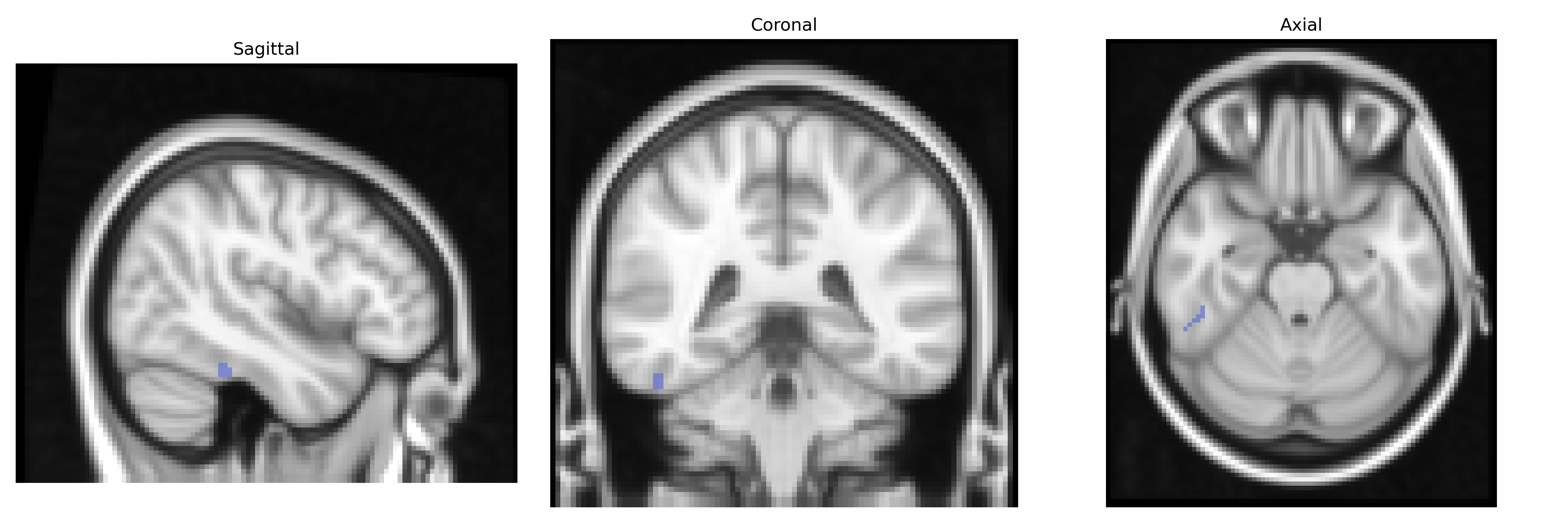
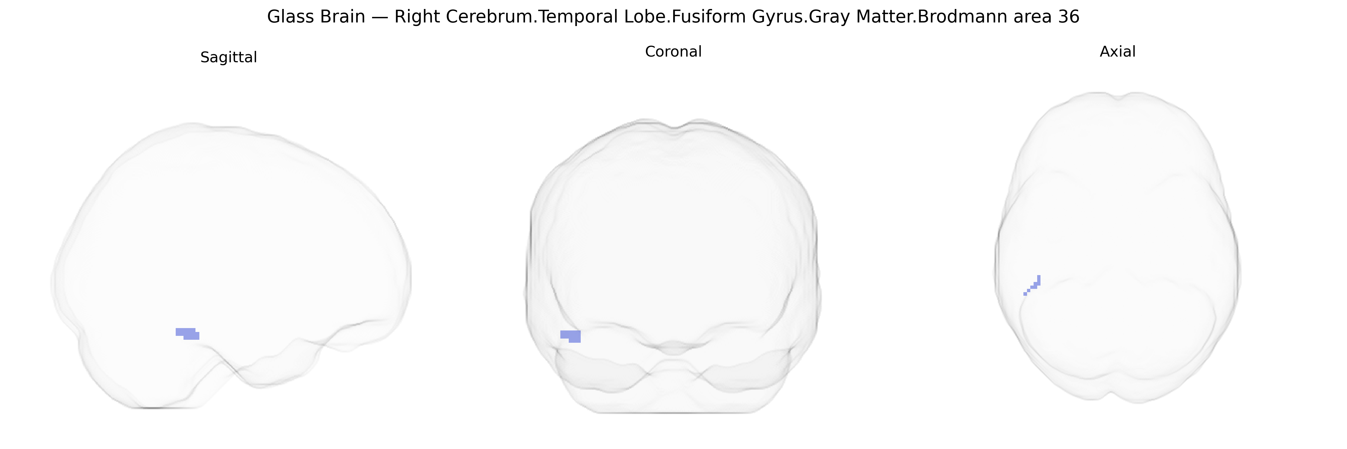
Bilateral Right Cerebrum.Temporal Lobe.Fusiform Gyrus.Gray Matter.Brodmann area 36 corresponds to a cytoarchitectonic subdivision of the parahippocampal/medial fusiform region within the temporal lobe, often associated with the ectorhinal cortex and bordering perirhinal cortex (BA35). This region participates in higher-order visual and multimodal processing, including object and face recognition, memory-related associative integration, and semantic processing, linking visual inputs with stored representations. Gray matter in BA36 contains layers of pyramidal and interneuron populations that project to and receive input from adjacent temporal, limbic, and prefrontal areas, supporting networks involved in recognition memory, familiarity judgments, and the encoding and retrieval of complex visual scenes and objects. There is no direct Wikipedia article for Brodmann area 36; see the related structure Parahippocampal gyrus.
Genetic associations involving the right fusiform gyrus gray matter in Brodmann area 36 (temporal lobe, as defined in the Talairach 2 mm atlas) largely emerge from imaging genetics and GWAS of brain structure and function rather than region-specific candidate studies. Variants in genes affecting synaptic plasticity, neurodevelopment, and cortical morphology—such as BDNF, DISC1, and several loci identified in large ENIGMA and UK Biobank GWAS—have been linked to fusiform cortical thickness and surface area, often shared across temporal and occipitotemporal regions. Fusiform gray matter and functional activation patterns in this area are implicated in face and object recognition, with genetic influences evidenced by heritability of fusiform activation during face processing and by GWAS signals related to social cognition and facial recognition ability, though most findings span bilateral fusiform regions without strict lateralization. Neurodevelopmental and psychiatric disorders, including autism spectrum disorder, schizophrenia, and major depression, show altered structure and connectivity in fusiform and adjacent BA36 cortex, with risk variants in genes such as CNTNAP2, NRXN1, and synaptic signaling loci contributing to these phenotypes; however, these are typically associated with wider temporal and limbic networks rather than anatomically isolated BA36. Rare variant and CNV studies (e.g., 22q11.2 deletion) also implicate fusiform and parahippocampal regions in visual and socio-emotional processing deficits. Overall, genetic findings support a polygenic influence on the morphology and function of BA36 fusiform cortex, embedded in broader temporal and ventral visual pathways, with no single gene uniquely specific to this bilateral right-hemisphere region.
Overview generated by GPT-4o (2026).
Region ID: 166
Hemisphere: bilateral
Atlas: Talairach labels 2mm
Full Quality Version: Download MP4
Full Quality Version: Download MP4
Wali Sidiqyar*, Gaurav Rudravaram*, Elyssa M. McMaster, Trent M. Schwartz, Adam M. Saunders, Kurt G. Schilling, Bennett A. Landman "Introducing SPINS: A Shared Public Visualization Library of Neuroanatomical Structures." Medical Imaging with Deep Learning- short paper
This resource is licensed under CC0 1.0 Universal (Public Domain).