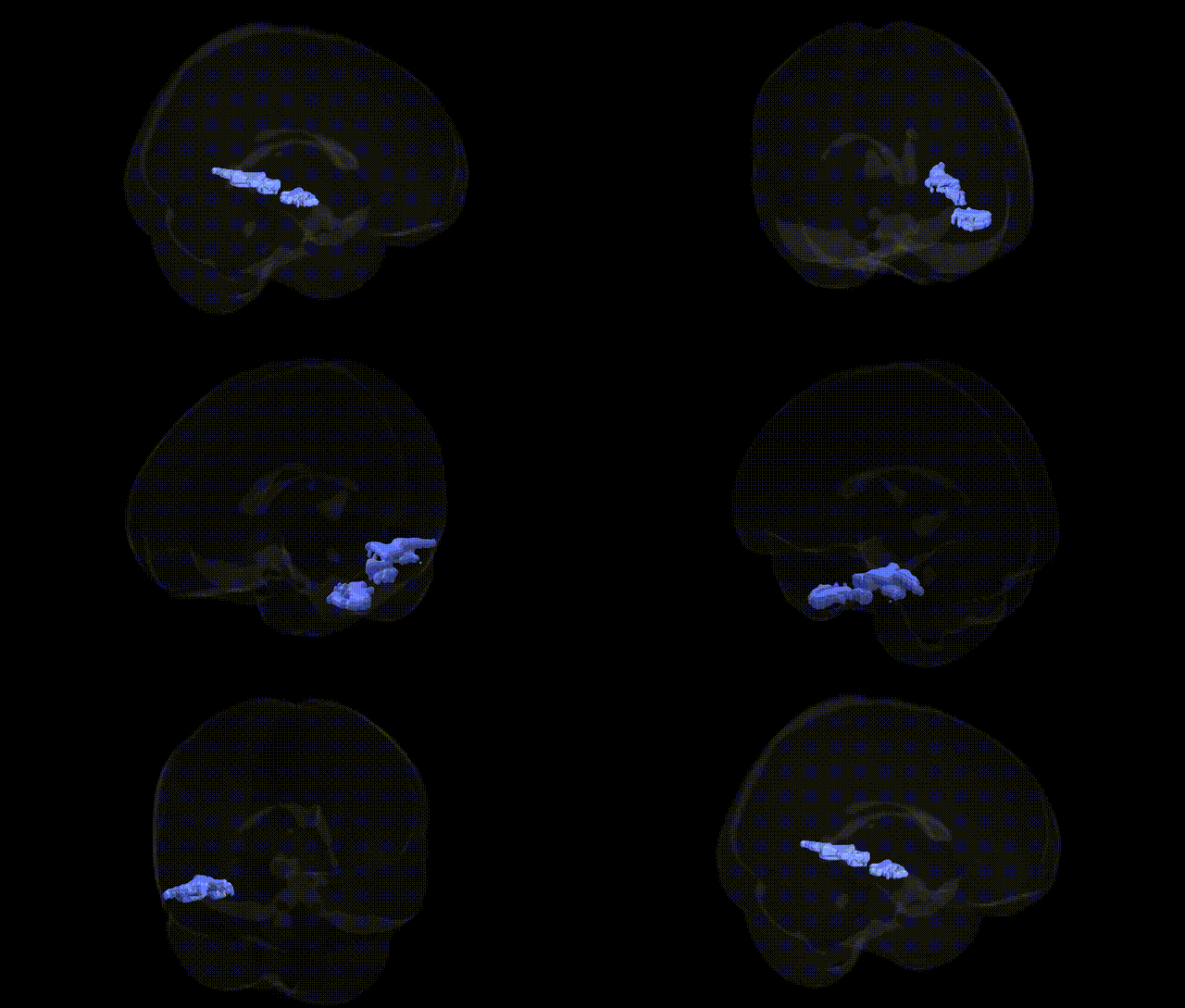
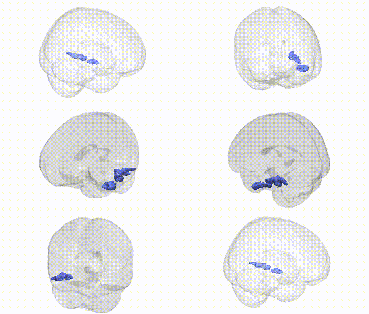
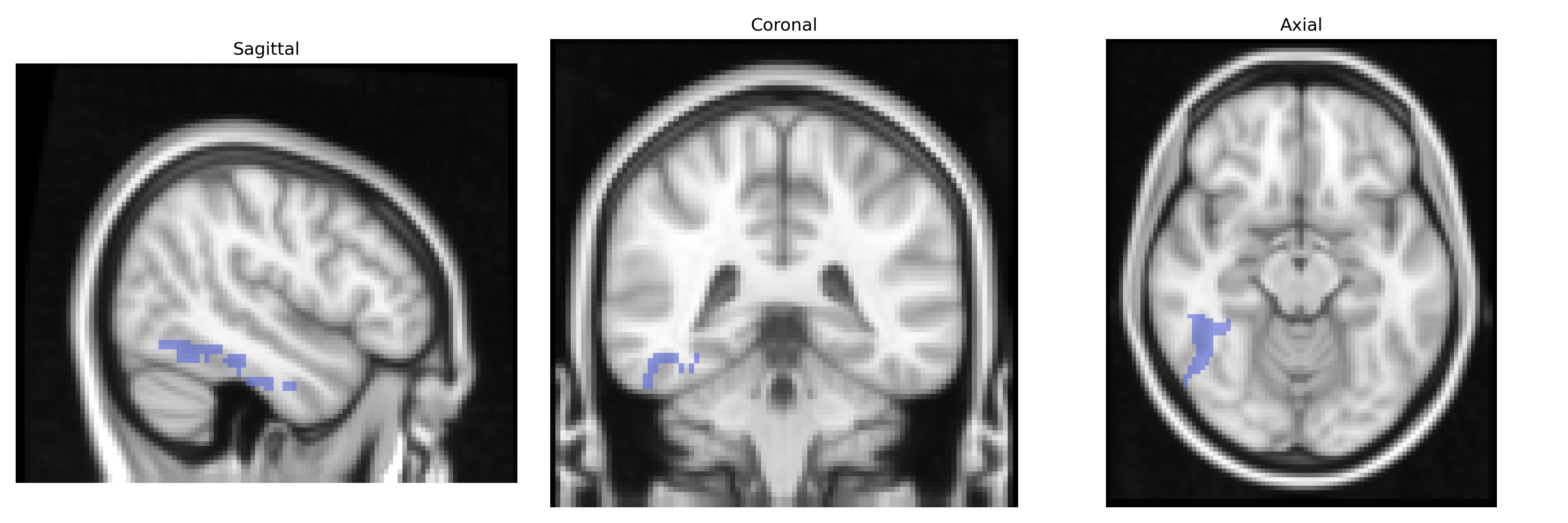
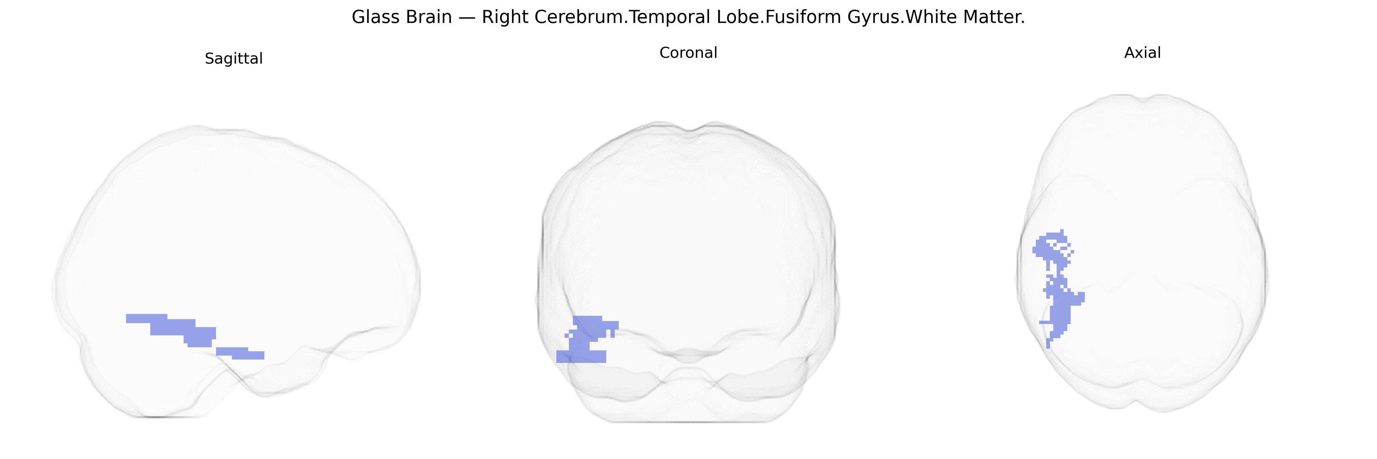
The bilateral Right Cerebrum.Temporal Lobe.Fusiform Gyrus.White Matter corresponds to the subcortical fiber pathways underlying the fusiform gyrus in the right temporal lobe, supporting communication between fusiform cortex and other temporal, occipital, and limbic regions. Functionally, this white matter region is crucial for the integrity of networks involved in high-level visual processing, including face and object recognition, as well as aspects of language and semantic integration through connections with adjacent temporal and occipitotemporal association areas. Damage or disconnection in this area can contribute to deficits such as visual agnosias or impairments in facial recognition (prosopagnosia), reflecting disruption of information transfer rather than primary cortical damage. There is no direct link for the specific white matter label, but it is closely related to the Fusiform gyrus.
The bilateral right temporal lobe fusiform gyrus white matter, as defined in the Talairach 2 mm atlas, has not typically been isolated as a unique target in genetic studies, but convergent evidence from GWAS and imaging-genetics work implicates genes and variants that influence fusiform structure, connectivity, and function in relation to face processing, reading, and social cognition. Polygenic scores for schizophrenia, autism spectrum disorder, and major depressive disorder have been associated with altered fusiform gray and white matter microstructure and volume, with notable contributions from risk loci in genes such as CACNA1C, DISC1, and common variants in the MHC region, which broadly modulate temporal and occipitotemporal connectivity. GWAS of cortical morphology (e.g., ENIGMA consortium studies) identify multiple loci (including variants near genes such as HMGA2, PAX8, and KIAA0586) associated with temporal and occipitotemporal cortical surface area and thickness, likely influencing underlying fusiform white matter tracts. Imaging-genetics studies of reading and dyslexia have linked variants in KIAA0319, DCDC2, and ROBO1 to altered integrity of temporo-occipital white matter pathways that encompass the fusiform region, consistent with its role in orthographic and visual word processing. In autism, risk variants in synaptic and neurodevelopmental genes (e.g., NRXN1, SHANK3, CNTNAP2) have been tied to atypical fusiform activation and connectivity in social and face-perception tasks, with diffusion MRI showing genotype-dependent differences in temporal lobe white matter organization. Although GWAS rarely report a single locus as specifically and exclusively tied to the right fusiform gyrus white matter, the region features repeatedly as a node within heritable structural and functional networks associated with neurodevelopmental disorders, psychosis, affective traits, cognitive performance, and reading ability, reflecting polygenic influences on temporal and occipitotemporal circuitry rather than discrete region-specific genetic effects.
Overview generated by GPT-4o (2026).
Region ID: 131
Hemisphere: bilateral
Atlas: Talairach labels 2mm
Full Quality Version: Download MP4
Full Quality Version: Download MP4
Wali Sidiqyar*, Gaurav Rudravaram*, Elyssa M. McMaster, Trent M. Schwartz, Adam M. Saunders, Kurt G. Schilling, Bennett A. Landman "Introducing SPINS: A Shared Public Visualization Library of Neuroanatomical Structures." Medical Imaging with Deep Learning- short paper
This resource is licensed under CC0 1.0 Universal (Public Domain).