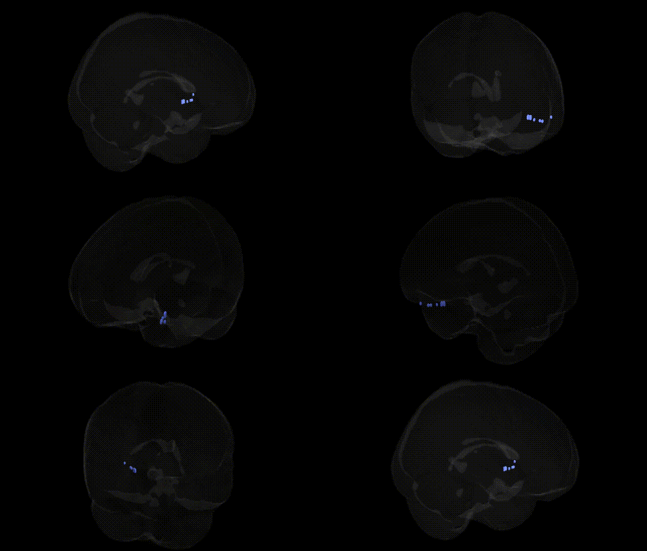
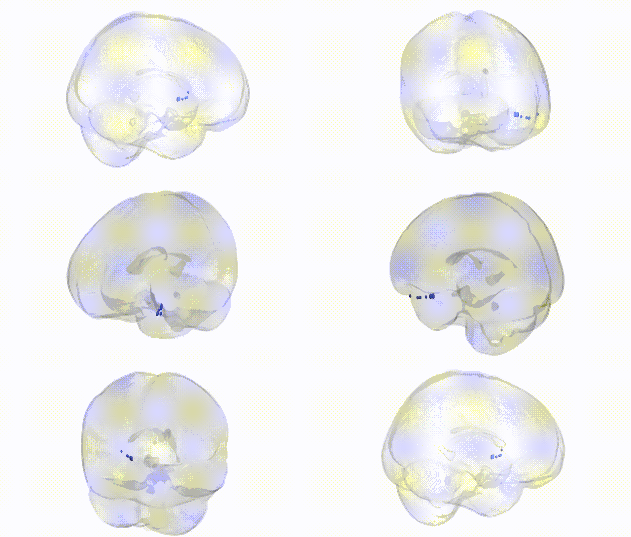
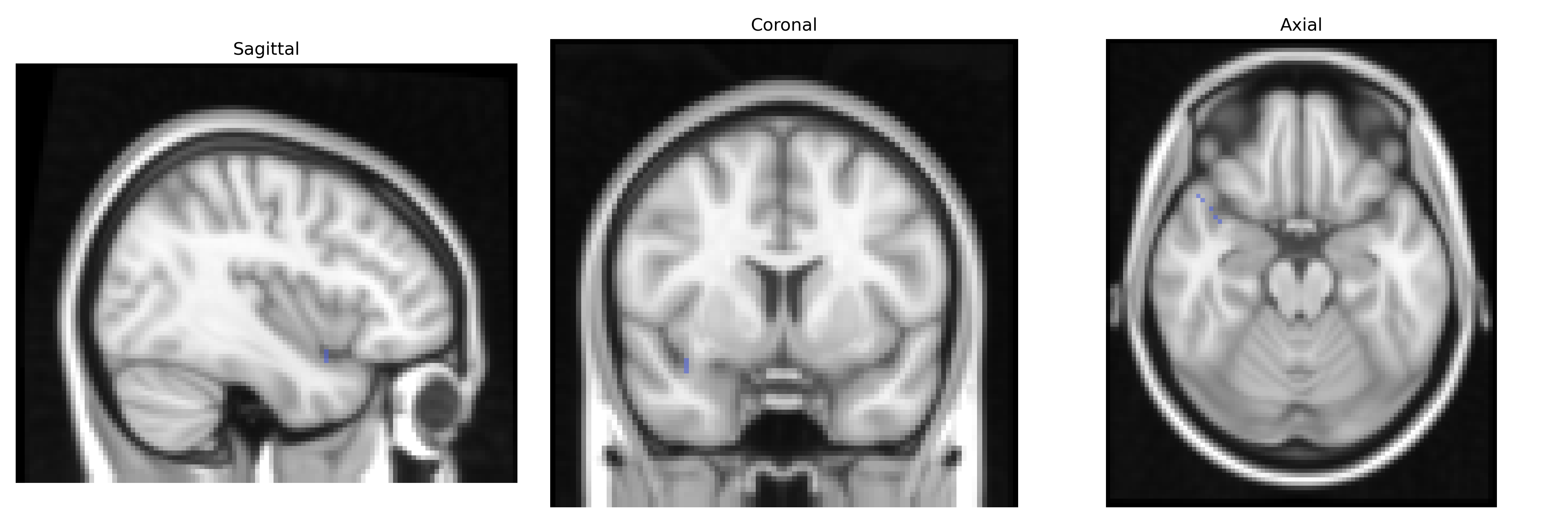
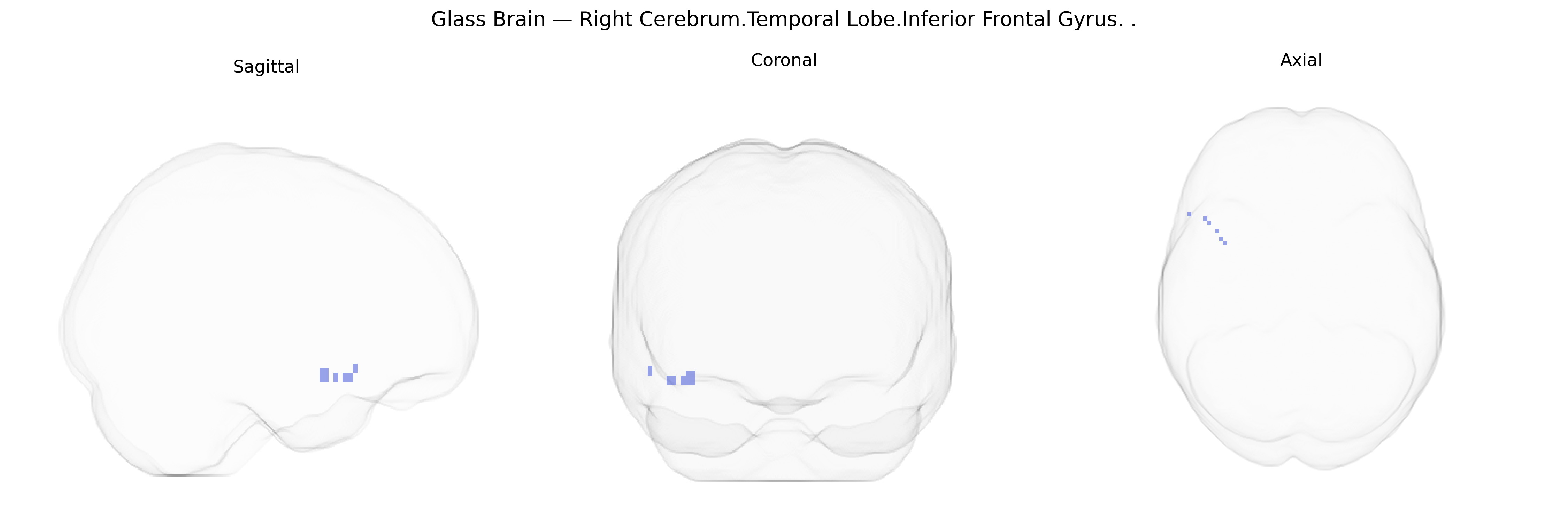
The bilateral Right Cerebrum.Temporal Lobe.Inferior Frontal Gyrus, as labeled in the Talairach 2 mm Atlas, reflects a border or mislabeling between the temporal lobe and the inferior frontal gyrus, a frontal lobe structure. The inferior frontal gyrus (IFG) is involved in language processing, speech production, cognitive control, and aspects of social cognition, with its pars opercularis and pars triangularis in the dominant hemisphere forming a core component of Broca’s area. Functionally, the IFG participates in syntactic processing, phonological working memory, response inhibition, and the selection among competing representations, while also contributing to the mirror neuron system and action understanding. Cytoarchitectonically, it lies anterior to the precentral gyrus and superior to the lateral sulcus, integrating inputs from temporal, parietal, and other frontal regions as part of distributed language and executive networks.
Inferior frontal gyrus
Genetic associations for the bilateral Right Cerebrum.Temporal Lobe.Inferior Frontal Gyrus region (as defined in the Talairach 2 mm atlas, broadly corresponding to inferior frontal and adjacent temporal cortices implicated in language, social cognition, and executive control) emerge mainly from imaging genetics and GWAS of brain structure and function rather than region-specific single-gene studies. Large GWAS of cortical morphology (e.g., ENIGMA, UK Biobank) have identified common variants near genes such as MAPT, KIAA0586, and others that influence inferior frontal and temporal cortical thickness and surface area, with downstream relevance to language and cognitive performance. Functional imaging‑genetic work has linked FOXP2 and CNTNAP2 variants to activation and connectivity in inferior frontal and temporal language circuits, especially during speech and phonological tasks. In psychiatric genetics, polygenic risk scores for schizophrenia, bipolar disorder, and major depression show associations with altered volume and activation in inferior frontal/temporal regions, and risk variants in CACNA1C, ZNF804A, and MIR137 have been associated with frontotemporal connectivity abnormalities. Autism spectrum disorder and social communication–related GWAS highlight genes such as NRXN1 and SHANK3 that affect temporo‑frontal networks, including inferior frontal areas engaged in social cognition and language. Neurodegenerative disease genetics (e.g., GRN, C9orf72, and MAPT variants) also show structural and functional effects in frontotemporal cortices, including inferior frontal regions, consistent with patterns of atrophy in frontotemporal dementia. Overall, genetic findings suggest that common and rare variation influencing synaptic function, neurodevelopment, and cortical plasticity contributes to interindividual differences in structure and function of the inferior frontal/temporal language–executive network, and these differences are implicated across language traits, general cognition, major psychiatric disorders, and frontotemporal neurodegeneration.
Overview generated by GPT-4o (2026).
Region ID: 229
Hemisphere: bilateral
Atlas: Talairach labels 2mm
Full Quality Version: Download MP4
Full Quality Version: Download MP4
Wali Sidiqyar*, Gaurav Rudravaram*, Elyssa M. McMaster, Trent M. Schwartz, Adam M. Saunders, Kurt G. Schilling, Bennett A. Landman "Introducing SPINS: A Shared Public Visualization Library of Neuroanatomical Structures." Medical Imaging with Deep Learning- short paper
This resource is licensed under CC0 1.0 Universal (Public Domain).