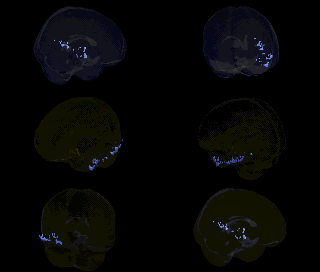
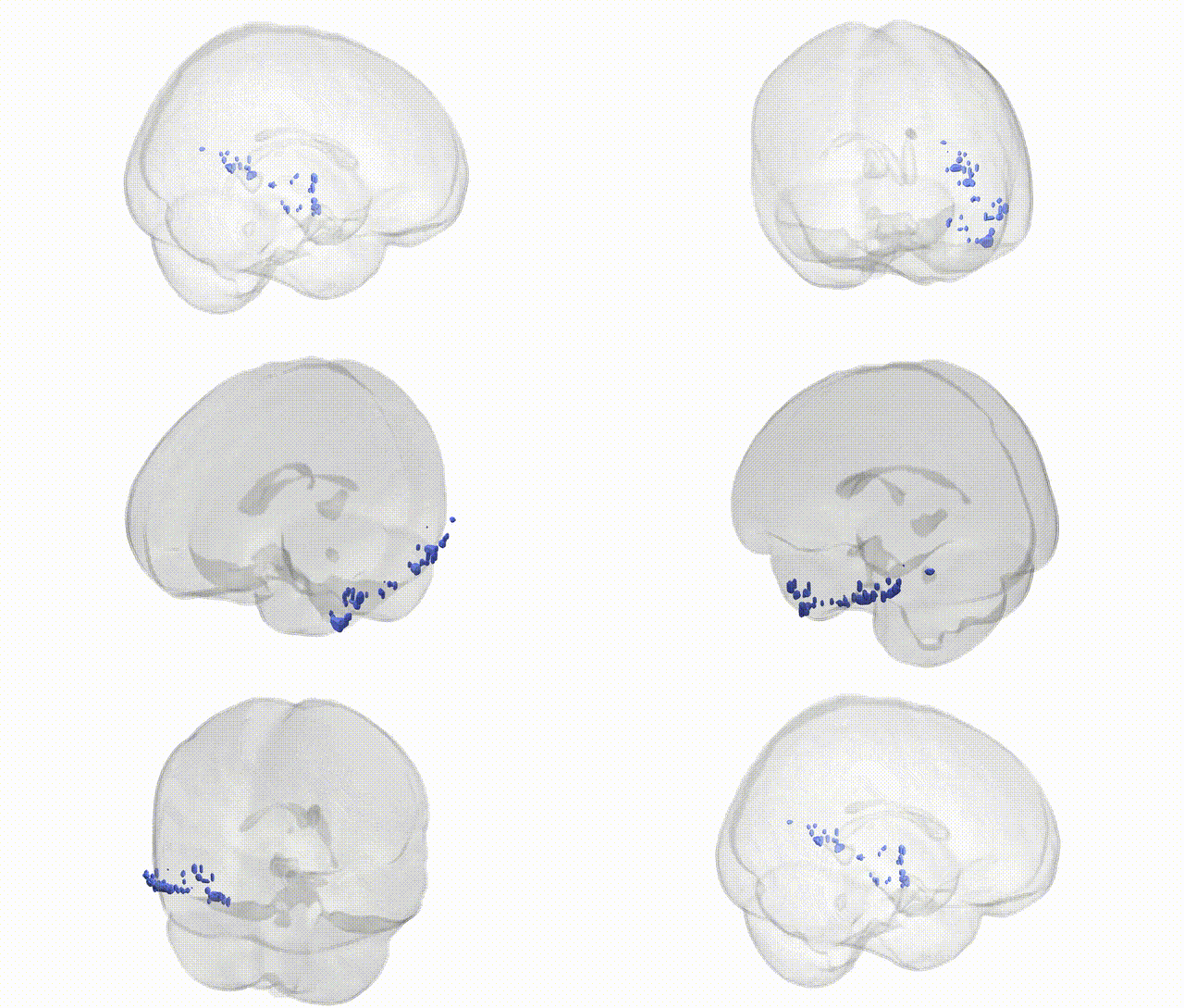
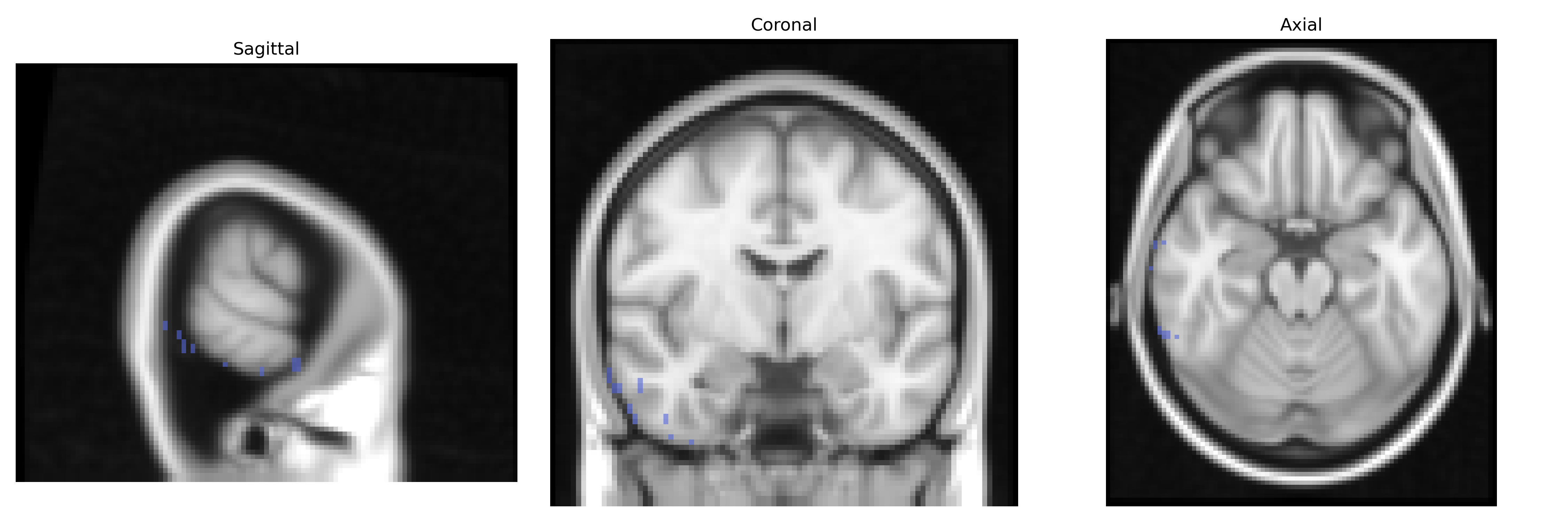
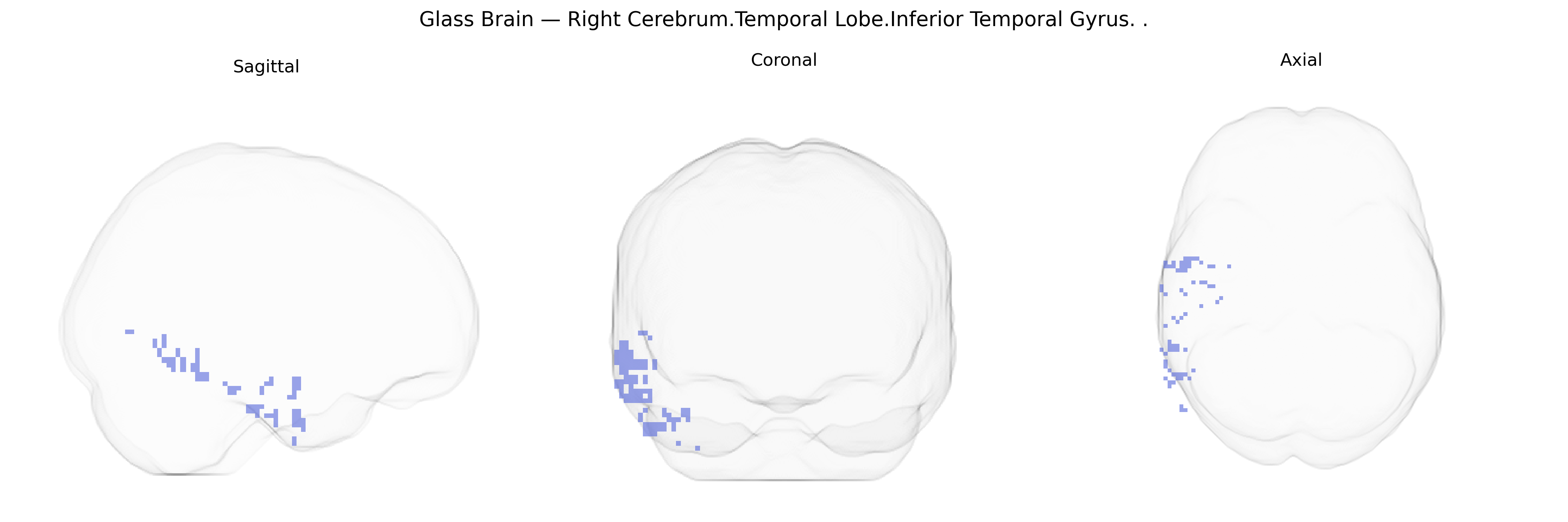
The bilateral Right Cerebrum.Temporal Lobe.Inferior Temporal Gyrus is a cortical region on the ventrolateral aspect of the temporal lobe, extending along the inferior temporal surface and involved primarily in high-level visual processing. It contributes to object recognition, visual form perception, and complex feature integration, forming part of the ventral “what” pathway that supports identification and categorization of visual stimuli. Neuronal populations in this gyrus respond to shapes, objects, and sometimes faces, and it has strong reciprocal connections with other temporal, occipital, and ventral visual stream areas. Functionally, it participates in semantic processing and may be implicated in disorders of visual recognition (agnosias) and certain aspects of language and memory when damaged. There is no direct link for the inferior temporal gyrus itself; a closely related structure is the Temporal lobe.
Genetic associations involving the inferior temporal gyrus, particularly in the right temporal lobe, arise largely from imaging‐genetics and GWAS studies that link specific variants to cortical thickness, surface morphology, and functional activation in this region rather than to the Talairach atlas label per se. Common variants in genes involved in synaptic development and plasticity (e.g., BDNF, NRG1, GRIN2B, DISC1) and in axonal guidance or neuronal migration (e.g., RELN, CNTNAP2) have been associated with structural and functional differences in inferior temporal and adjacent fusiform cortex, which supports high-level visual processing, face perception, and semantic memory. Neuropsychiatric GWAS and imaging genetics work in schizophrenia, autism spectrum disorder, major depressive disorder, and bipolar disorder have repeatedly implicated temporal lobe structures, with polygenic risk scores for these disorders showing correlations with reduced cortical thickness, altered gyrification, or atypical activation in inferior temporal regions. In Alzheimer’s disease and other dementias, GWAS-identified risk variants in APOE, CLU, PICALM, and BIN1 are associated with atrophy patterns that include the temporal neocortex, and tau and amyloid burden in temporal regions has been linked to genetic risk profiles. Large-scale ENIGMA consortium analyses have identified multiple loci (e.g., in genes related to neurodevelopment and cell adhesion) that modulate temporal lobe cortical thickness and surface area, some of which overlap with GWAS hits for cognitive performance, educational attainment, and general intelligence, suggesting that genetic influences on the inferior temporal gyrus contribute to variability in visual cognition and semantic processing as well as vulnerability to psychiatric and neurodegenerative disorders, even though no single gene is uniquely or specifically tied to the bilateral right inferior temporal gyrus in isolation.
Overview generated by GPT-4o (2026).
Region ID: 12
Hemisphere: bilateral
Atlas: Talairach labels 2mm
Full Quality Version: Download MP4
Full Quality Version: Download MP4
Wali Sidiqyar*, Gaurav Rudravaram*, Elyssa M. McMaster, Trent M. Schwartz, Adam M. Saunders, Kurt G. Schilling, Bennett A. Landman "Introducing SPINS: A Shared Public Visualization Library of Neuroanatomical Structures." Medical Imaging with Deep Learning- short paper
This resource is licensed under CC0 1.0 Universal (Public Domain).