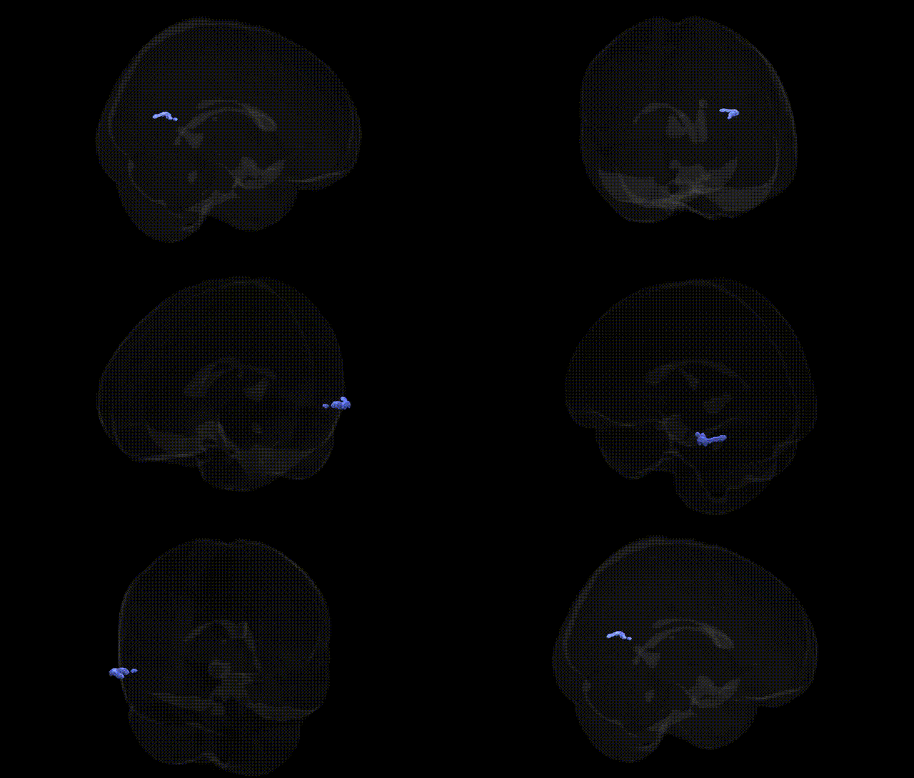
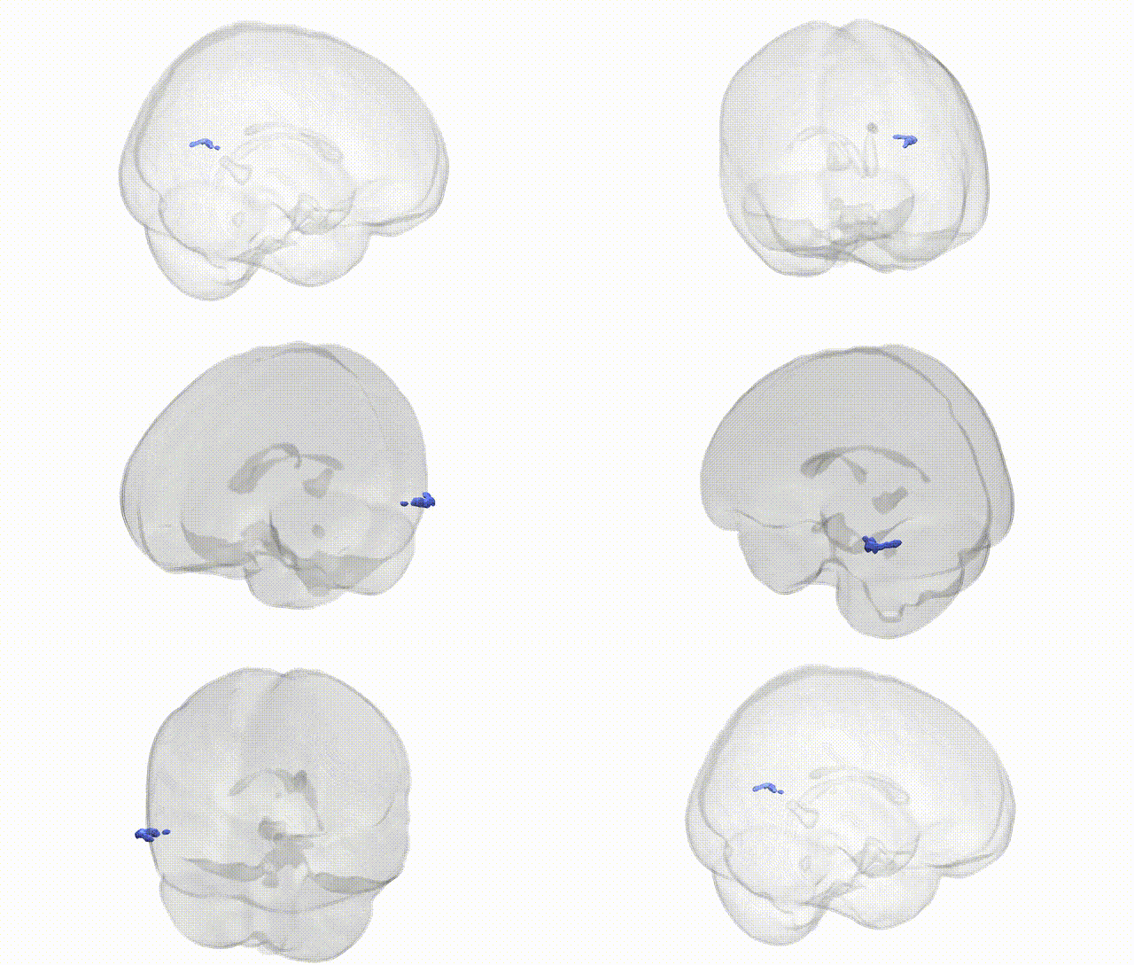
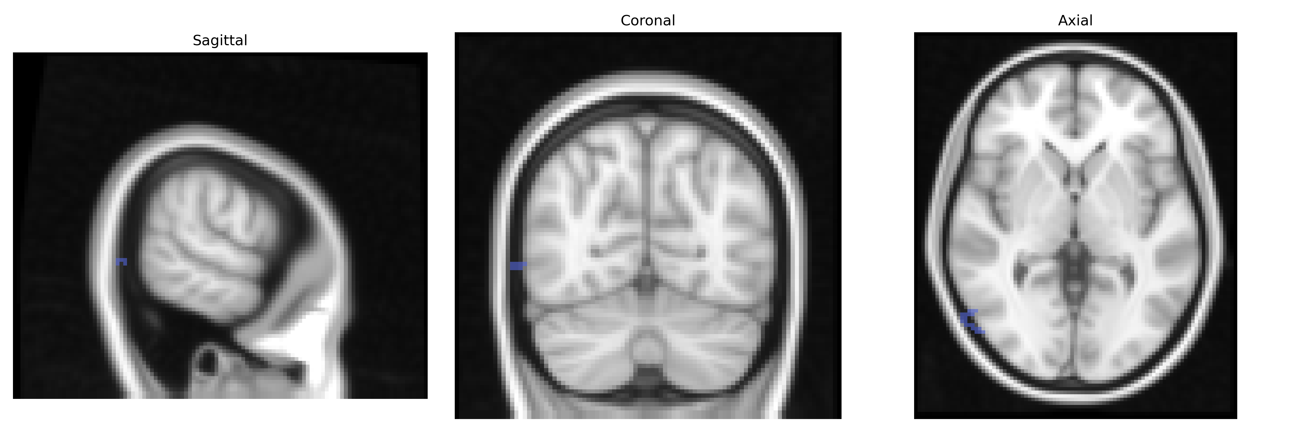
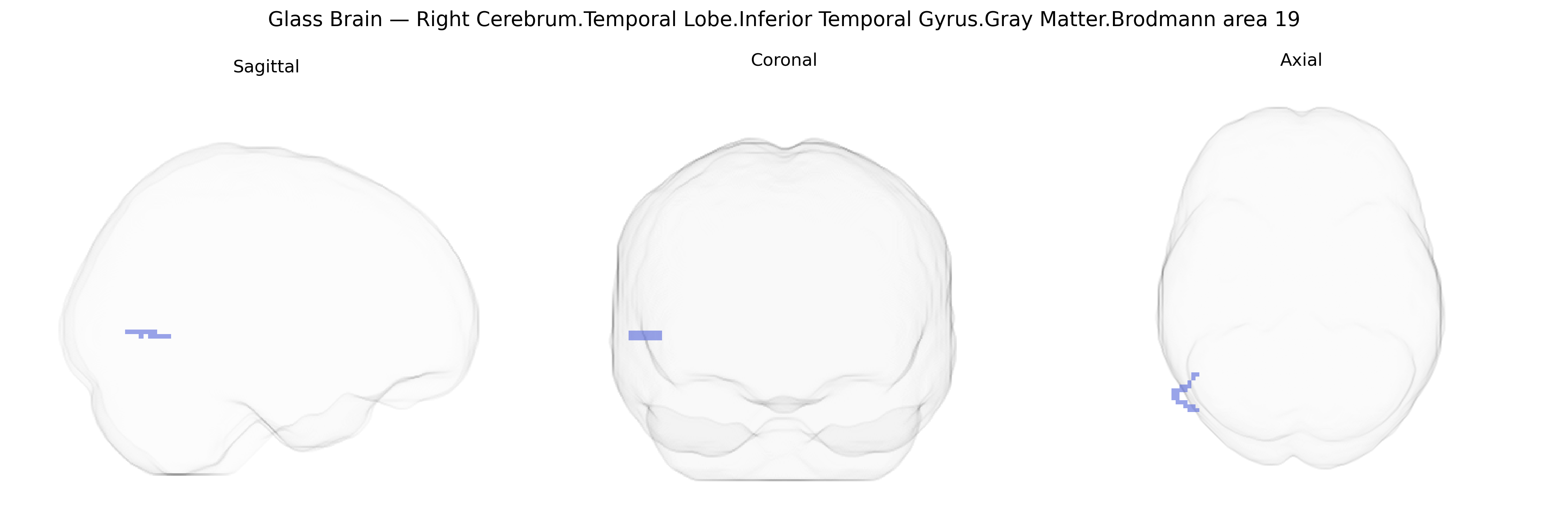
The bilateral Right Cerebrum.Temporal Lobe.Inferior Temporal Gyrus.Gray Matter.Brodmann area 19 corresponds to portions of the ventral visual association cortex that extend into the inferior temporal region, integrating complex visual information with higher-order perceptual and mnemonic processes. Brodmann area 19 is classically part of the extrastriate visual cortex and participates in the analysis of form, color, and motion, contributing to object recognition and visual pattern processing through reciprocal connections with primary visual cortex (BA17), secondary visual cortex (BA18), and temporal lobe structures involved in visual memory. Neurons in this region exhibit relatively large receptive fields and hierarchical feature selectivity, supporting transformation of basic visual inputs into more abstract representations that can be linked to semantic knowledge and recognition. There is no direct link for “Right Cerebrum.Temporal Lobe.Inferior Temporal Gyrus.Gray Matter.Brodmann area 19”; a closely related structure is Brodmann area 19.
The bilateral inferior temporal gyrus gray matter in Brodmann area 19, a higher-order visual association region, has been implicated in genetic studies primarily through imaging genetics and GWAS of brain structure, cognition, and neuropsychiatric risk rather than through region-specific single-gene findings. Large-scale GWAS of cortical thickness and surface area (e.g., ENIGMA, UK Biobank) have identified polygenic influences on temporal and occipital association cortices, with loci near genes involved in neurodevelopment (such as PAX6, KIAA0586, and variants in Wnt and synaptic pathways) contributing to variability in inferior temporal and BA19 morphology, though effects are highly distributed and not specific to this exact Talairach-defined parcel. Functionally, BA19 is engaged in high-level visual processing, object recognition, and aspects of reading, and genetic studies of developmental dyslexia and reading ability (implicating genes such as DCDC2, KIAA0319, and ROBO1) have reported structural and functional alterations in posterior temporal–occipital regions that overlap BA19, suggesting that risk alleles for language and reading disorders may partly act through development of this circuitry. GWAS and candidate-gene imaging studies in schizophrenia, bipolar disorder, and major depression (highlighting genes such as ZNF804A, CACNA1C, and variants in complement and synaptic genes) have shown associations with temporal and occipital cortical volumes and connectivity, including inferior temporal regions, consistent with the role of BA19 in visual cognition, social perception, and hallucination-related processing. In autism spectrum disorder, common and rare variants affecting synaptic, chromatin-remodeling, and neurodevelopmental genes (e.g., NRXN1, SHANK3, CHD8) have been linked to altered temporal–occipital cortical thickness and gyrification, and several imaging–genetics analyses report that polygenic risk for autism correlates with atypical development of lateral temporal and visual association areas encompassing BA19. Overall, genetic influences on this region are highly polygenic, shared across large-scale networks, and most robustly detected through GWAS of global cortical measures, cognitive traits (such as intelligence and reading), and neuropsychiatric disorders, with convergent evidence that variation in neurodevelopmental, synaptic, and plasticity-related genes shapes structure and function in inferior temporal BA19.
Overview generated by GPT-4o (2026).
Region ID: 480
Hemisphere: bilateral
Atlas: Talairach labels 2mm
Full Quality Version: Download MP4
Full Quality Version: Download MP4
Wali Sidiqyar*, Gaurav Rudravaram*, Elyssa M. McMaster, Trent M. Schwartz, Adam M. Saunders, Kurt G. Schilling, Bennett A. Landman "Introducing SPINS: A Shared Public Visualization Library of Neuroanatomical Structures." Medical Imaging with Deep Learning- short paper
This resource is licensed under CC0 1.0 Universal (Public Domain).