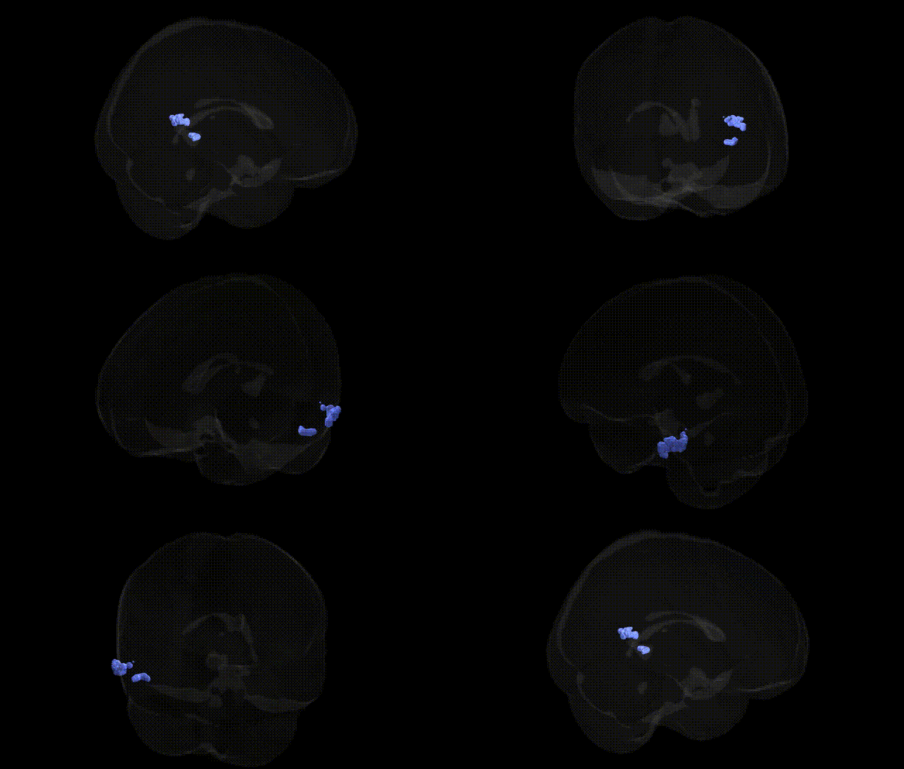
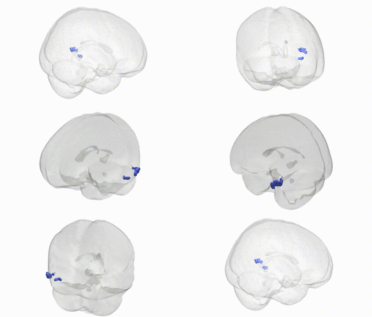
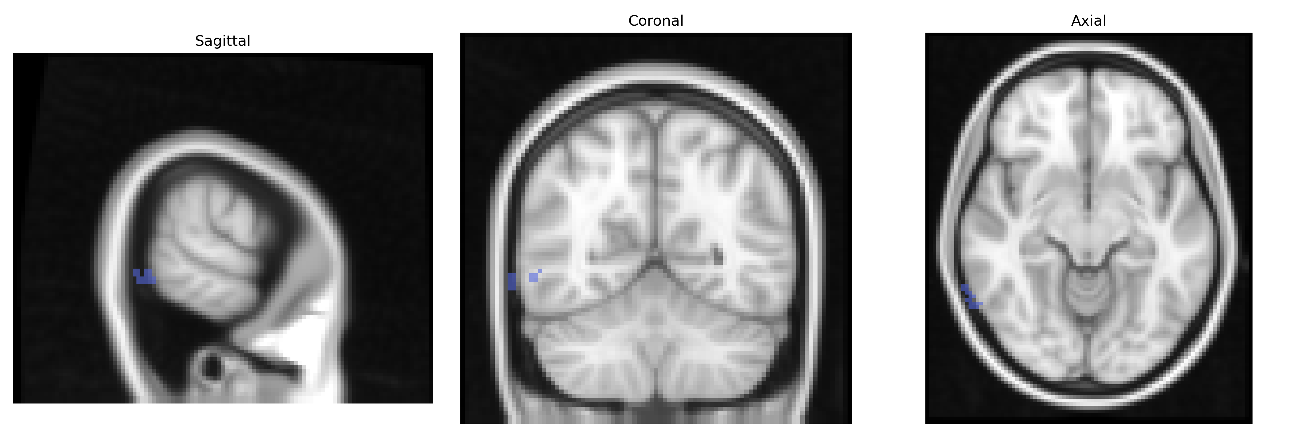
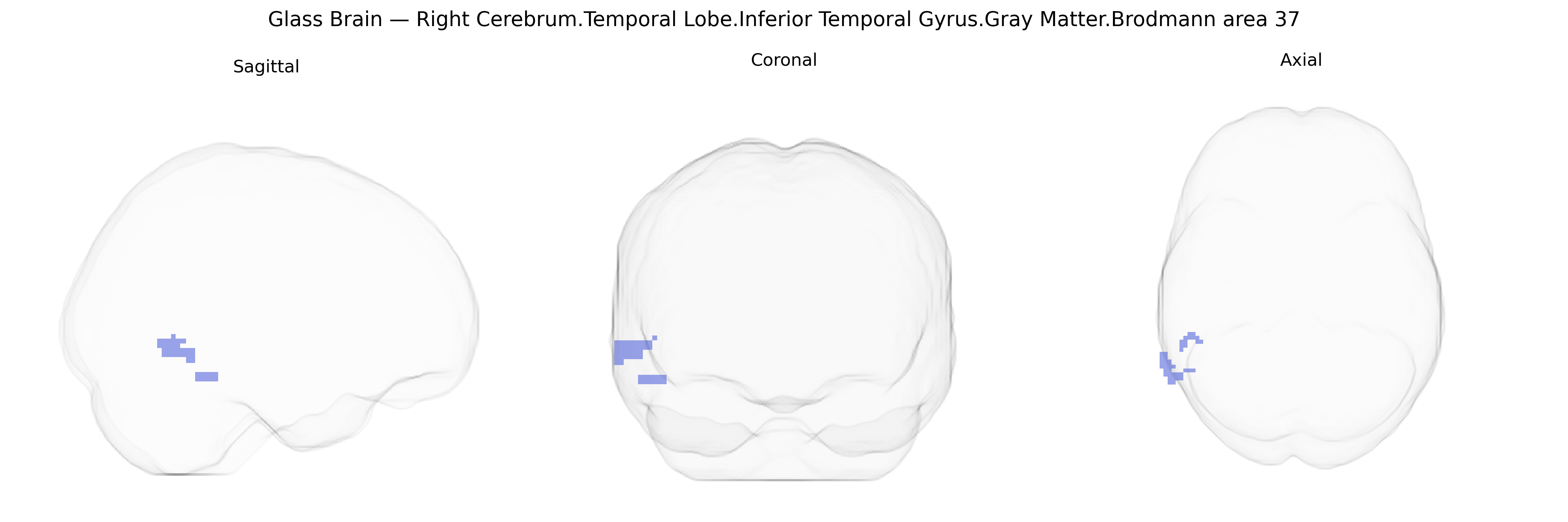
The bilateral Right Cerebrum.Temporal Lobe.Inferior Temporal Gyrus.Gray Matter.Brodmann area 37 corresponds primarily to the fusiform and adjacent inferior temporal regions implicated in high-level visual processing, particularly object and face recognition, and the integration of complex visual features with semantic memory. Cytoarchitectonically defined as Brodmann area 37, this cortex lies in the posterior inferior temporal lobe, bordering occipitotemporal regions and receiving dense input from ventral visual stream areas, thus participating in category-specific recognition (e.g., words, faces, and other complex stimuli) and multimodal association functions. Functionally, BA37 contributes to the mapping of visual percepts onto stored representations, supporting lexical retrieval and semantic processing, and is often associated with deficits such as visual agnosia or category-specific recognition impairments when damaged.
Fusiform gyrus
The bilateral right inferior temporal gyrus (Brodmann area 37) gray matter, as defined in the Talairach 2 mm atlas, lies within the ventral visual stream and overlaps the fusiform gyrus, a region repeatedly implicated in genetic studies of cognition, language, and neuropsychiatric disease. GWAS and imaging‑genetics work on cortical surface area and thickness have shown that variation in BA37/ventral temporal cortex structure is influenced by common variants in loci involved in neurodevelopment and synaptic function (for example, genes near microtubule and axon guidance pathways), although specific high‑effect variants are rare and effects are highly polygenic. The broader BA37/fusiform territory has been associated with reading and face‑processing abilities, with multiple genetic studies of dyslexia and developmental language disorder implicating genes such as DCDC2, KIAA0319, and ROBO1, which affect temporal and occipitotemporal cortical development and connectivity. In autism spectrum disorder, rare and common variants in synaptic and chromatin‑regulating genes (e.g., SHANK3, NRXN1, CHD8) are linked to altered fusiform and inferior temporal gyrus volume and activation patterns, correlating with social‑perceptual deficits. Schizophrenia and bipolar disorder GWAS—highly polygenic and enriched for neuronal and synaptic genes—have been associated at the imaging level with reduced gray matter and altered functional activity in inferior temporal and fusiform regions, suggesting that shared genetic risk impacts this ventral temporal network. In Alzheimer’s disease and other dementias, risk variants such as APOE ε4 and loci affecting amyloid and tau (e.g., CLU, BIN1, PTK2B) have been linked in imaging‑genetics studies to early atrophy and hypometabolism in temporal lobe association cortices including BA37, reflecting vulnerability of higher‑order visual and semantic processing regions. Overall, genetic influences on this region are distributed across many loci identified in large GWAS of cortical morphology, cognition, and major neuropsychiatric and neurodegenerative disorders, with BA37 emerging as a key structural and functional node whose variability partially reflects underlying polygenic risk.
Overview generated by GPT-4o (2026).
Region ID: 214
Hemisphere: bilateral
Atlas: Talairach labels 2mm
Full Quality Version: Download MP4
Full Quality Version: Download MP4
Wali Sidiqyar*, Gaurav Rudravaram*, Elyssa M. McMaster, Trent M. Schwartz, Adam M. Saunders, Kurt G. Schilling, Bennett A. Landman "Introducing SPINS: A Shared Public Visualization Library of Neuroanatomical Structures." Medical Imaging with Deep Learning- short paper
This resource is licensed under CC0 1.0 Universal (Public Domain).