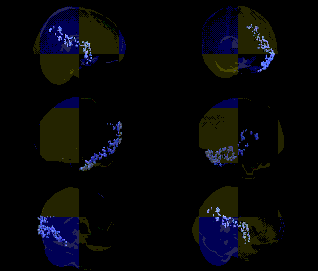
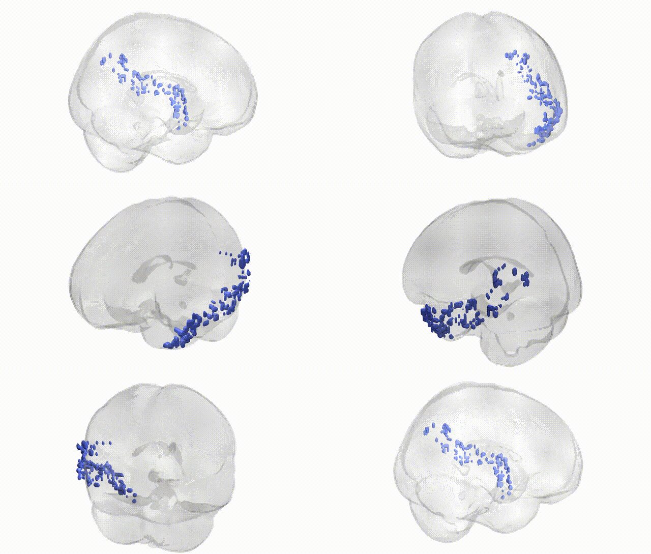
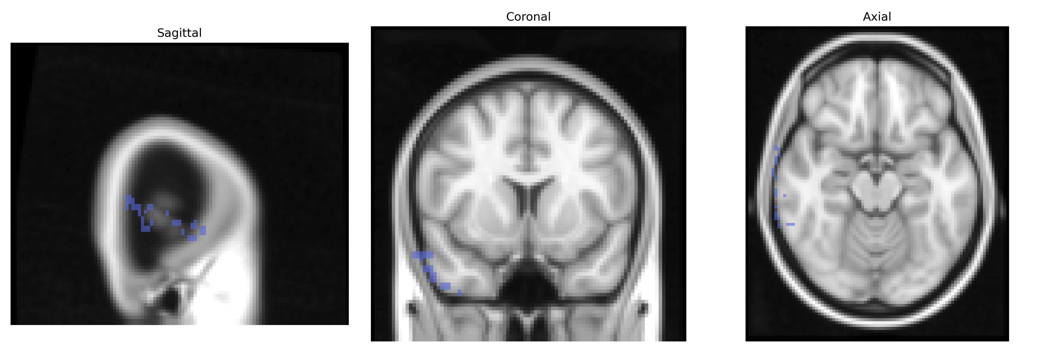
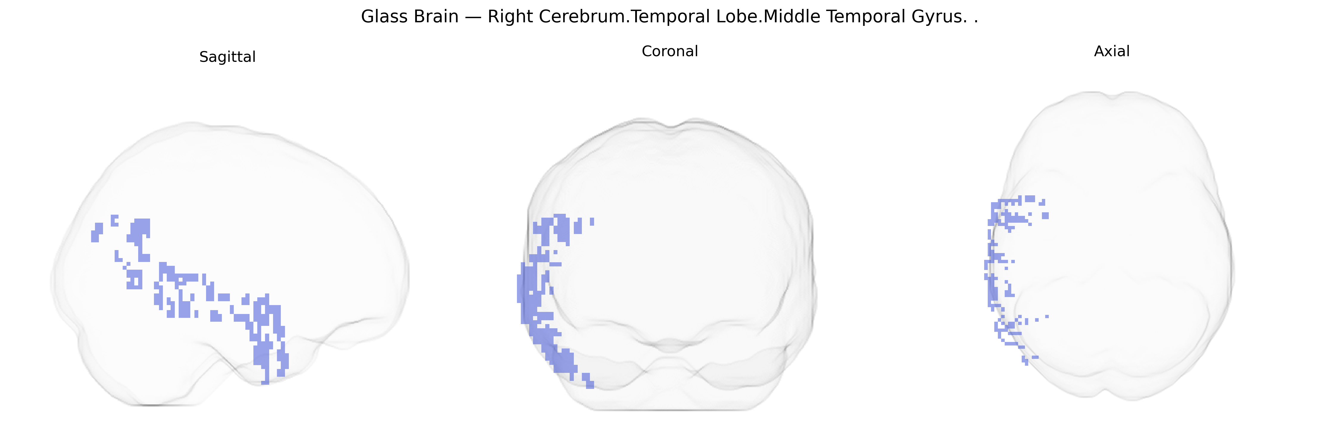
The bilateral right cerebrum temporal lobe middle temporal gyrus is a neocortical region on the lateral aspect of the right temporal lobe, extending rostrocaudally between the superior and inferior temporal sulci. It is primarily associated with higher-order auditory and language processing, semantic memory, and aspects of social cognition, including the interpretation of biological motion and facial expressions. Functionally, it participates in the ventral visual stream and multimodal association networks, integrating auditory, visual, and linguistic information, and contributes to semantic comprehension and lexical retrieval, particularly in the dominant hemisphere. Pathology involving the right middle temporal gyrus has been implicated in deficits in social perception, semantic processing, and certain forms of aphasia and temporal lobe epilepsy.
Middle temporal gyrus
The bilateral right middle temporal gyrus (MTG) of the temporal lobe, as defined in the Talairach 2 mm atlas, has been implicated in genetic studies primarily through imaging–genetics and GWAS of brain structure and function rather than single-gene associations specific to this exact parcel. Large-scale GWAS of cortical morphology (e.g., ENIGMA and UK Biobank cohorts) have identified common variants near genes involved in neurodevelopment, synaptic function, and axon guidance—such as MEF2C, DCC, and genes in glutamatergic pathways—associated with temporal lobe cortical thickness and surface area, which often include or overlap the MTG. Imaging–genetics work in language and social cognition frequently implicates temporal lobe regions including MTG, with polygenic risk for schizophrenia, autism spectrum disorder, and major depressive disorder linked to altered temporal lobe activation and connectivity, although these signals are typically regional rather than parcel-specific. GWAS of reading ability, language-related traits, and educational attainment also show polygenic influences on temporal cortical structure and functional activation, with MTG contributing to semantic and language networks that are sensitive to these genetic effects. Additionally, risk alleles for Alzheimer’s disease (such as APOE ε4 and variants in CLU, PICALM, and BIN1) are associated with temporal lobe atrophy and hypometabolism, which may encompass the MTG, and polygenic risk scores for neurodegenerative and psychiatric disorders have been correlated with temporal lobe measures in structural and functional MRI, suggesting that genetic liability for these conditions partly manifests through this region’s anatomy and network integration, even though no single gene has been uniquely or consistently tied specifically to the bilateral right MTG in current GWAS catalogs.
Overview generated by GPT-4o (2026).
Region ID: 30
Hemisphere: bilateral
Atlas: Talairach labels 2mm
Full Quality Version: Download MP4
Full Quality Version: Download MP4
Wali Sidiqyar*, Gaurav Rudravaram*, Elyssa M. McMaster, Trent M. Schwartz, Adam M. Saunders, Kurt G. Schilling, Bennett A. Landman "Introducing SPINS: A Shared Public Visualization Library of Neuroanatomical Structures." Medical Imaging with Deep Learning- short paper
This resource is licensed under CC0 1.0 Universal (Public Domain).