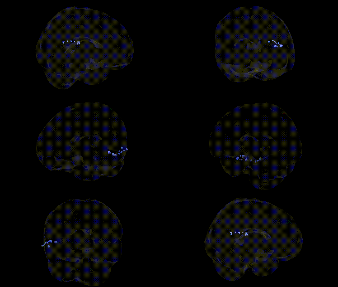
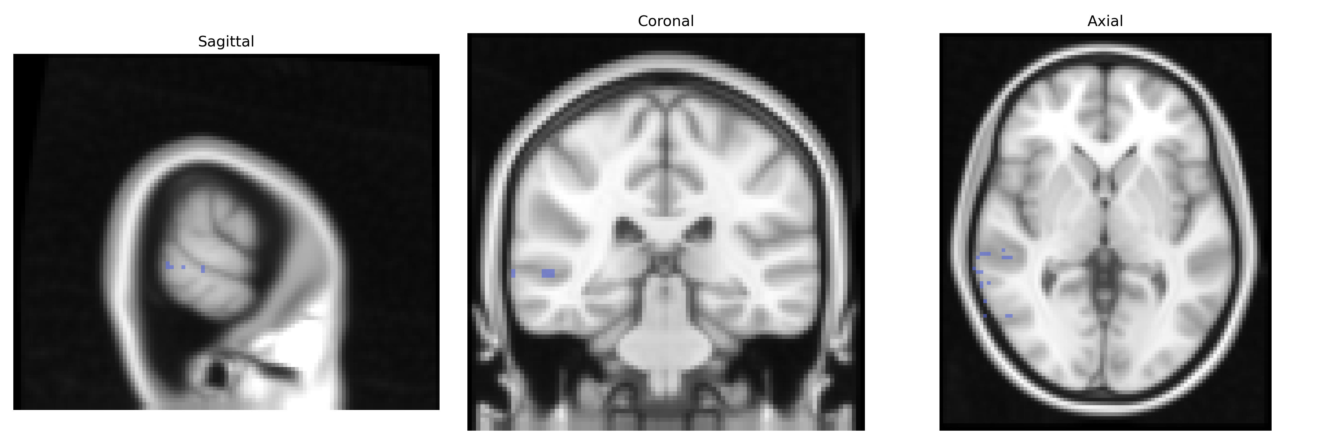
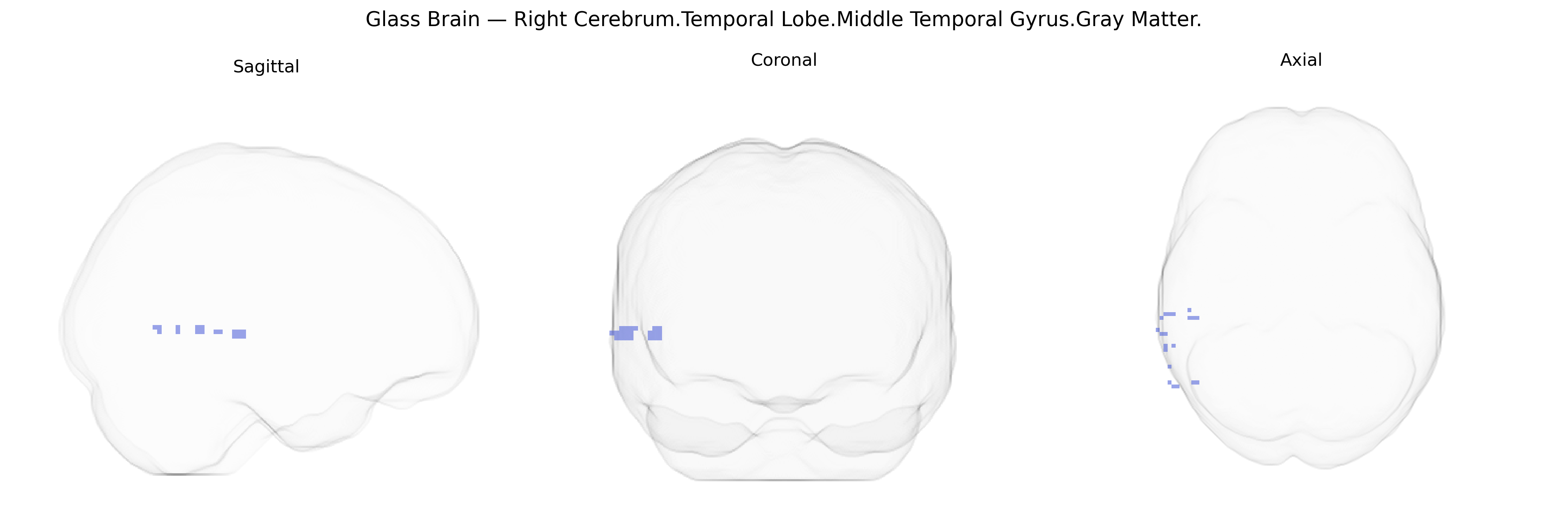
The bilateral Right Cerebrum.Temporal Lobe.Middle Temporal Gyrus.Gray Matter corresponds to cortical gray matter within the right middle temporal gyrus, a longitudinally oriented region on the lateral aspect of the temporal lobe involved in multimodal associative processing. This area participates in language and semantic processing, lexical retrieval, and aspects of social cognition, including the interpretation of complex visual and auditory stimuli. It is also implicated in higher-order visual perception (such as motion and object recognition), integration of auditory information, and contributes to networks supporting comprehension and conceptual knowledge. Cytoarchitectonically, the middle temporal gyrus includes portions of association cortex that are interconnected with frontal, parietal, and occipital regions, forming part of large-scale semantic and default-mode networks.
The bilateral right middle temporal gyrus gray matter (as defined in the Talairach 2 mm atlas) is frequently implicated in imaging genetics and GWAS of brain structure and function, particularly in relation to cortical thickness and surface area, with variants in genes involved in neurodevelopment and synaptic plasticity (such as BDNF, CNTNAP2, and DISC1) showing associations with structural measures in this temporal region. Large-scale GWAS of MRI-derived cortical phenotypes have identified loci affecting temporal lobe morphology that overlap with risk variants for schizophrenia, bipolar disorder, major depressive disorder, and autism spectrum disorder, reflecting shared genetic influences on temporal lobe architecture and psychiatric vulnerability. Middle temporal gyrus structure and activation are also linked to genetic risk for language and reading-related traits (e.g., FOXP2, DCDC2, KIAA0319), social cognition and theory-of-mind performance, and face and semantic processing, with polygenic scores for cognitive ability and educational attainment showing correlations with temporal lobe volumes. In Alzheimer’s disease and frontotemporal dementia, risk variants in genes such as APOE, MAPT, and GRN have been associated with patterns of temporal lobe atrophy that include the middle temporal gyrus, while imaging-genetic studies in epilepsy and temporal lobe epilepsy highlight genetic contributions (e.g., in sodium channel and GABA-related genes) to temporal cortical abnormalities. Collectively, GWAS and candidate-gene studies converge on the middle temporal gyrus as a key node where genetic variation influencing neurodevelopment, language and higher cognition, and susceptibility to major neuropsychiatric and neurodegenerative disorders manifests in altered gray matter structure and function.
Overview generated by GPT-4o (2026).
Region ID: 555
Hemisphere: bilateral
Atlas: Talairach labels 2mm
Full Quality Version: Download MP4
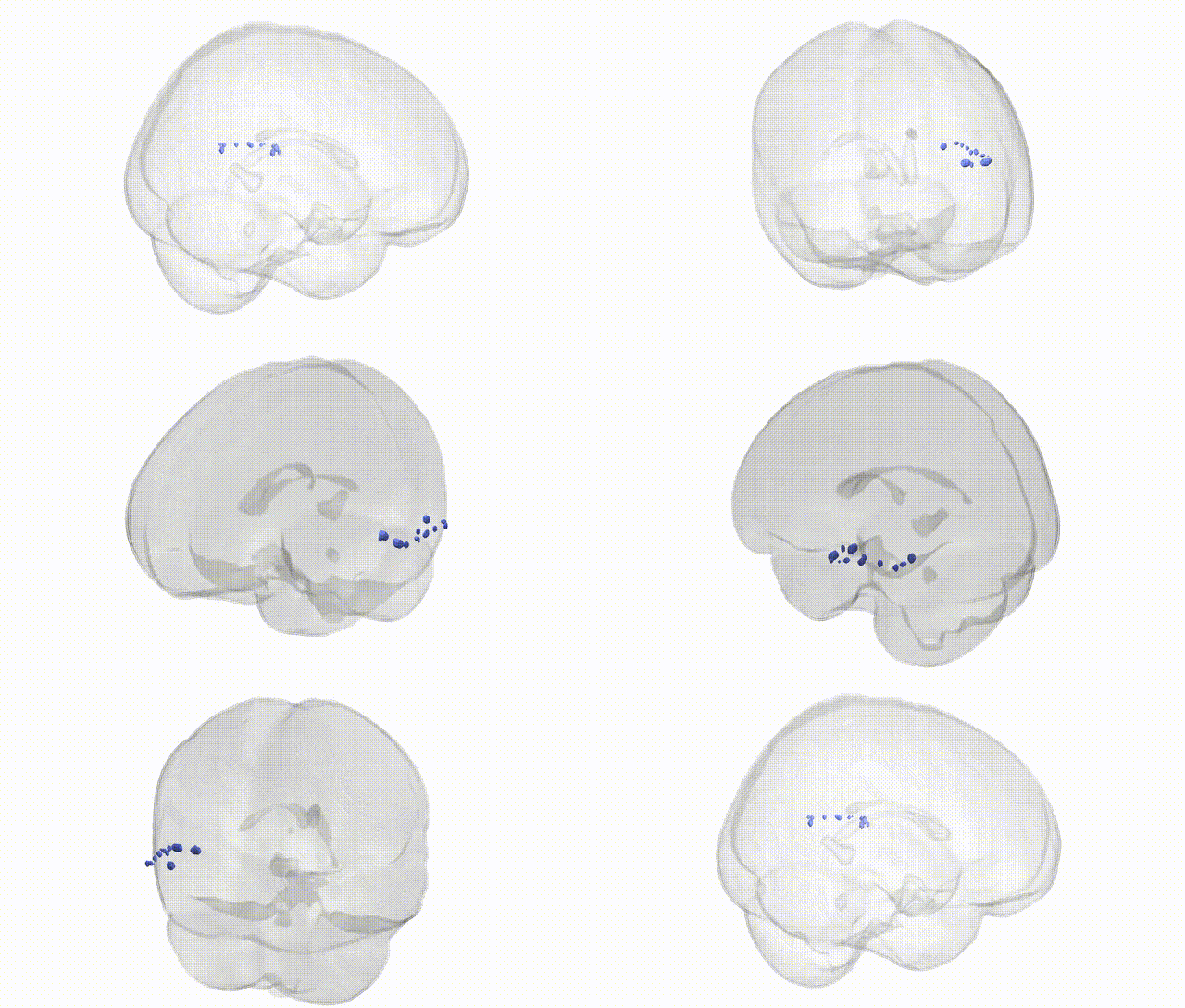
Full Quality Version: Download MP4
Wali Sidiqyar*, Gaurav Rudravaram*, Elyssa M. McMaster, Trent M. Schwartz, Adam M. Saunders, Kurt G. Schilling, Bennett A. Landman "Introducing SPINS: A Shared Public Visualization Library of Neuroanatomical Structures." Medical Imaging with Deep Learning- short paper
This resource is licensed under CC0 1.0 Universal (Public Domain).