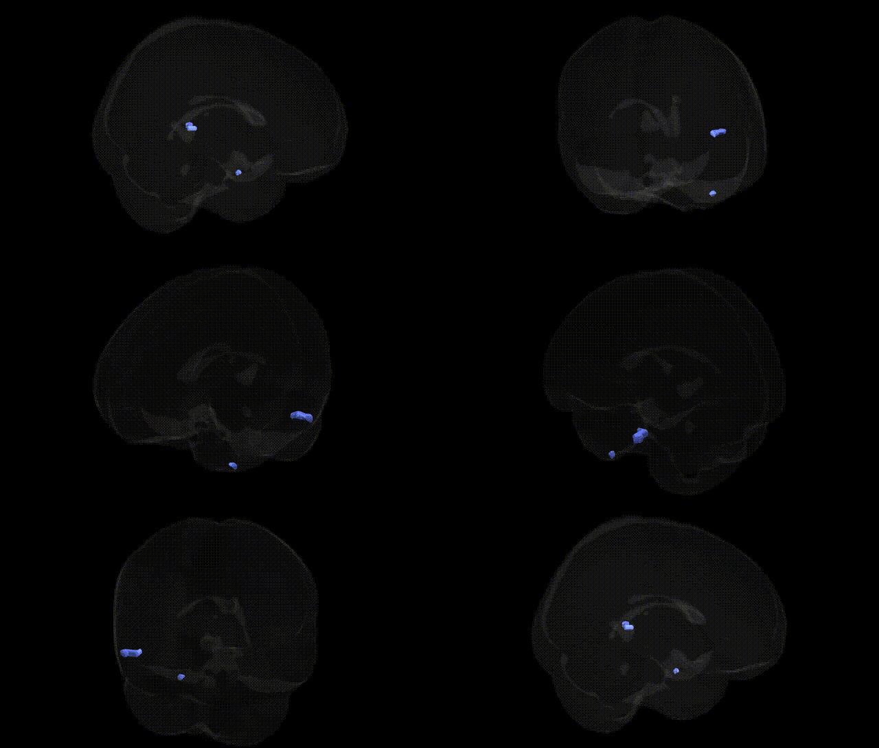
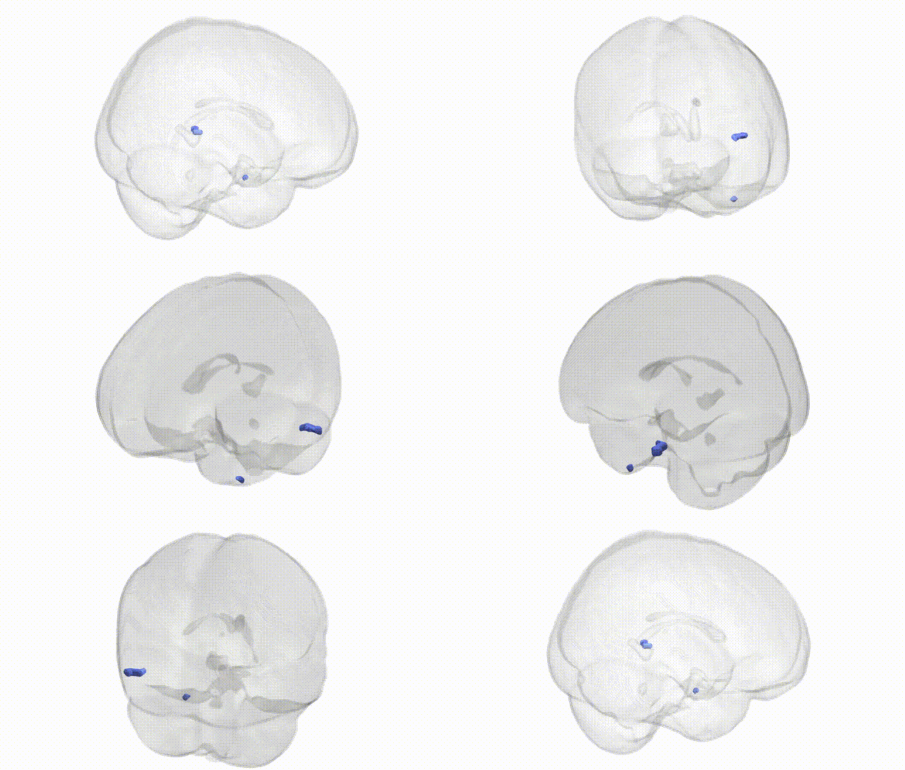
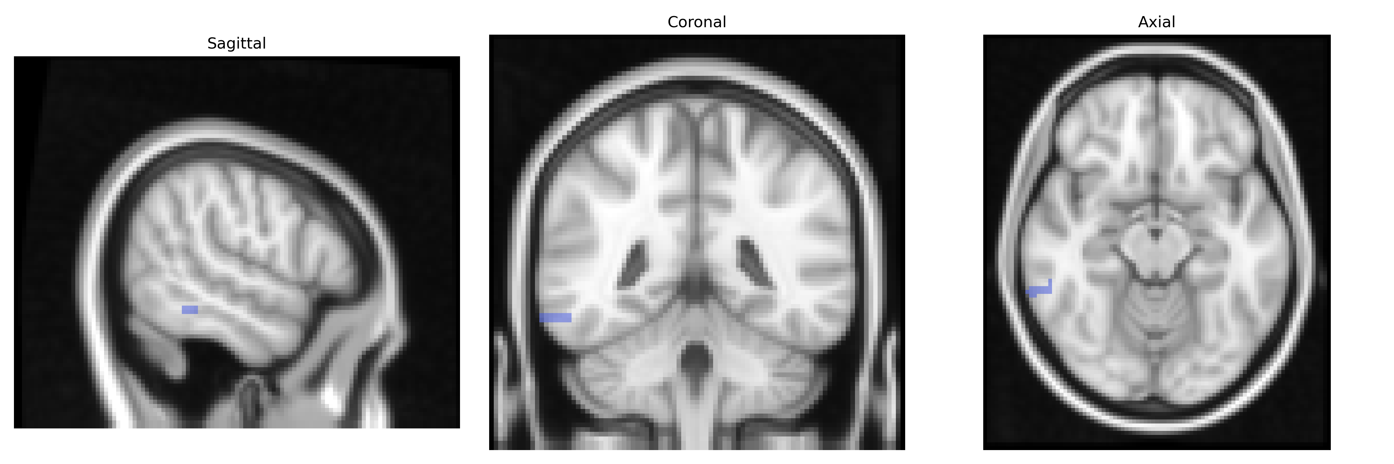
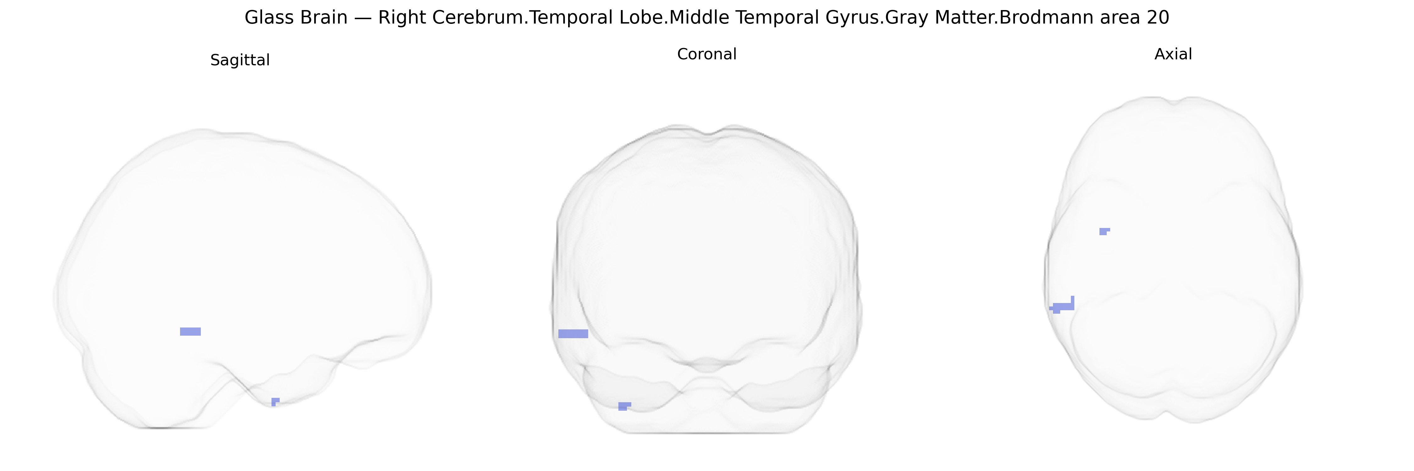
The bilateral Right Cerebrum.Temporal Lobe.Middle Temporal Gyrus.Gray Matter.Brodmann area 20 corresponds to ventral portions of the middle temporal gyrus in the inferior temporal cortex, implicated in high-level visual processing and semantic aspects of object and face recognition. Cytoarchitectonically defined as Brodmann area 20, this region contains association cortex that integrates visual inputs from occipito-temporal pathways with multimodal information to support recognition, categorization, and memory-related functions. It is part of the broader temporal lobe network involved in language comprehension, conceptual knowledge, and visual-semantic integration, and has reciprocal connections with other temporal association areas, the fusiform gyrus, and limbic structures. There is no direct link for Brodmann area 20; a related structure is the Middle temporal gyrus.
The bilateral right middle temporal gyrus gray matter in Brodmann area 20, as defined in the Talairach 2 mm atlas, has been implicated in genetic studies primarily through its roles in language, semantic memory, social cognition, and higher-order visual processing. GWAS and imaging-genetics work indicate that common variants influencing cortical thickness and surface area in temporal regions, including BA20, are enriched in neurodevelopmental and synaptic genes (for example, pathways involving glutamatergic signaling and axon guidance), and polygenic scores for educational attainment, general cognitive ability, and reading-related skills show associations with structural and functional measures in the middle and inferior temporal gyri. Genetic risk for autism spectrum disorder, schizophrenia, and major depressive disorder has been linked to altered temporal lobe morphology and connectivity, with several large consortia (e.g., ENIGMA) reporting that higher polygenic risk for these disorders correlates with volume or thickness differences in ventral temporal cortex encompassing BA20. Additionally, variants in genes such as CNTNAP2, FOXP2, and others involved in language and social communication have been associated with functional activation and connectivity changes in temporal regions overlapping BA20 during speech, semantic, and face-processing tasks. In neurodegenerative disease, APOE ε4 and other Alzheimer’s disease risk loci have been connected to atrophy and hypometabolism in temporal neocortex regions including BA20, consistent with the role of this area in semantic memory and object recognition.
Overview generated by GPT-4o (2026).
Region ID: 29
Hemisphere: bilateral
Atlas: Talairach labels 2mm
Full Quality Version: Download MP4
Full Quality Version: Download MP4
Wali Sidiqyar*, Gaurav Rudravaram*, Elyssa M. McMaster, Trent M. Schwartz, Adam M. Saunders, Kurt G. Schilling, Bennett A. Landman "Introducing SPINS: A Shared Public Visualization Library of Neuroanatomical Structures." Medical Imaging with Deep Learning- short paper
This resource is licensed under CC0 1.0 Universal (Public Domain).